Copyright
©The Author(s) 2022.
World J Stem Cells. Aug 26, 2022; 14(8): 616-632
Published online Aug 26, 2022. doi: 10.4252/wjsc.v14.i8.616
Published online Aug 26, 2022. doi: 10.4252/wjsc.v14.i8.616
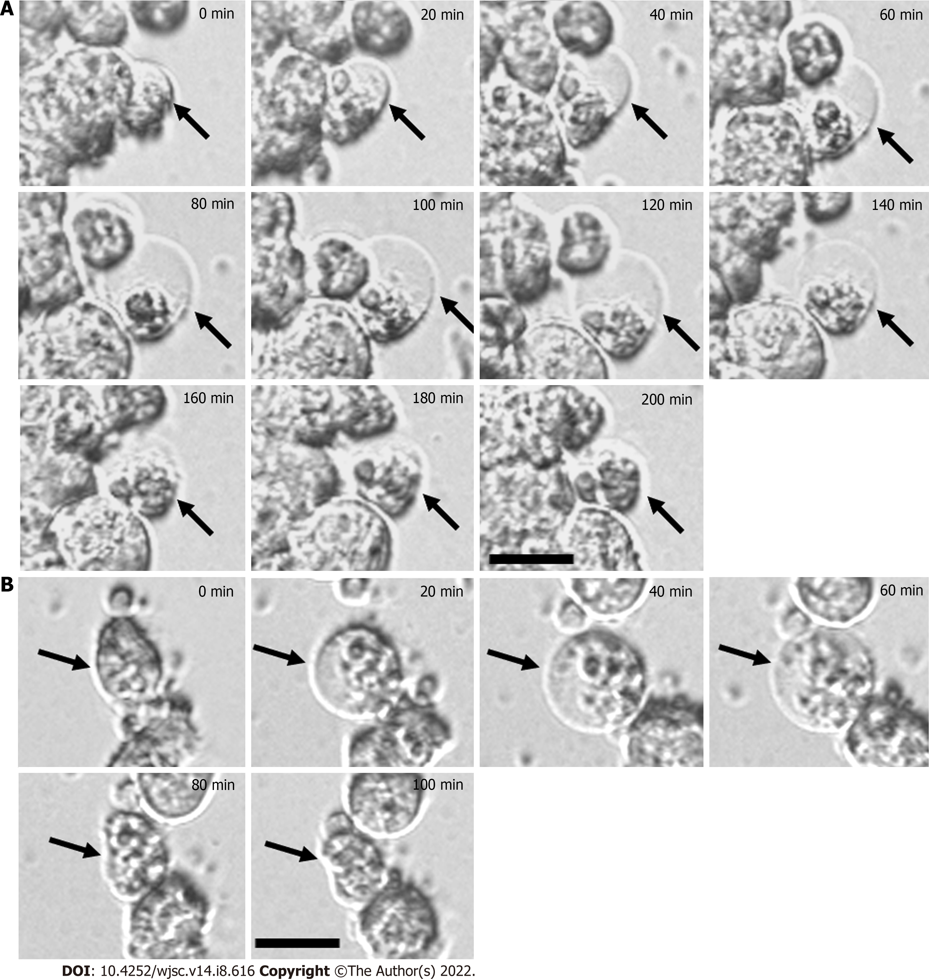
Figure 2 Time-lapse images showing the dynamic process of bubble formation for mesenchymal stem cells cultured on chitosan-hyaluronan or polyvinyl alcohol substrates.
A: Chitosan-hyaluronan; B: Polyvinyl alcohol. All images were collected after seeding of mesenchymal stem cells for 30 min. The process of bubble formation and disappearance was recorded by time-lapse phase contrast microscope. The dynamic changes of bubble structure were indicated by black arrows. The timeline in the upper right corner represents the initial snapshot of the immature bubble counted from the beginning (0 min to 200 min). Scale bar: 20 μm.
- Citation: Wong CW, Han HW, Hsu SH. Changes of cell membrane fluidity for mesenchymal stem cell spheroids on biomaterial surfaces. World J Stem Cells 2022; 14(8): 616-632
- URL: https://www.wjgnet.com/1948-0210/full/v14/i8/616.htm
- DOI: https://dx.doi.org/10.4252/wjsc.v14.i8.616


