Copyright
©The Author(s) 2022.
World J Stem Cells. Aug 26, 2022; 14(8): 616-632
Published online Aug 26, 2022. doi: 10.4252/wjsc.v14.i8.616
Published online Aug 26, 2022. doi: 10.4252/wjsc.v14.i8.616
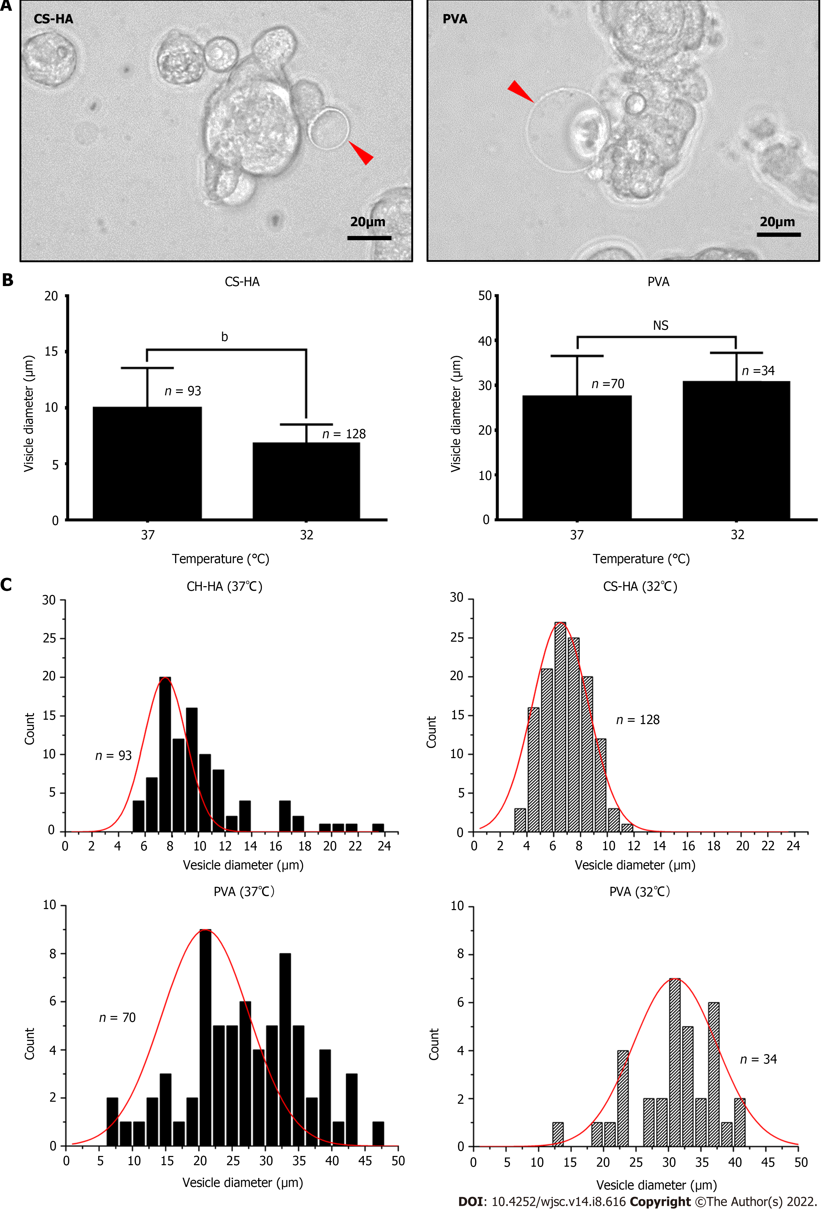
Figure 1 The morphologies of vesicle-like bubbles on the cell membrane of the spheroids of mesenchymal stem cells cultured on chitosan-hyaluronan or polyvinyl alcohol substrates.
A: Representative spheroids from cells cultured on chitosan-hyaluronan (CS-HA) substrates for 15 h are shown. The vesicle-like bubbles are indicated by red arrows. The scale bar represent 20 μm; B: The average diameter of the vesicle-like bubbles was measured and compared between different temperatures. Statistical significance was tested using the GraphPad Prim unpaired t-test. bP < 0.01; C: The frequency distribution histogram of vesicle-like bubbles diameter on the cell membrane for mesenchymal stem cells cultured on CS-HA or polyvinyl alcohol substrates at 37 °C or 32 °C. The frequency distribution histogram was obtained through the Gauss-amp fitting curve. n: Number of measured vesicle-like bubbles; PVA: Polyvinyl alcohol; MSCs: Mesenchymal stem cells; NS: Non-significant differences; CS: Chitosan; CS-HA: CS-hyaluronan.
- Citation: Wong CW, Han HW, Hsu SH. Changes of cell membrane fluidity for mesenchymal stem cell spheroids on biomaterial surfaces. World J Stem Cells 2022; 14(8): 616-632
- URL: https://www.wjgnet.com/1948-0210/full/v14/i8/616.htm
- DOI: https://dx.doi.org/10.4252/wjsc.v14.i8.616


