Copyright
©The Author(s) 2022.
World J Stem Cells. Aug 26, 2022; 14(8): 599-615
Published online Aug 26, 2022. doi: 10.4252/wjsc.v14.i8.599
Published online Aug 26, 2022. doi: 10.4252/wjsc.v14.i8.599
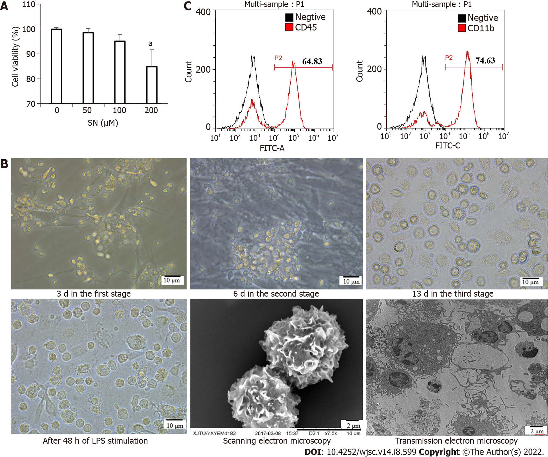
Figure 2 Morphological changes from induced pluripotent stem cells to induced pluripotent stem cells-dendritic cells.
A: The optimal concentration of sinomenine (SN) was 100 μM; B: Morphological changes from induced pluripotent stem cells (iPSCs) to iPSCs-dendritic cells (DCs) at 3 d in the first stage; iPSC-derived hematopoietic cells at 6 d in the second stage; and iPSC-derived imDCs at 13 d in the third stage under the influence of SN. After 48 h of lipopolysaccharide stimulation, the morphology of SN-iPSCs-DCs in phase contrast microscopy, scanning electron microscopy and transmission electron microscopy; C: Expression of the cell surface markers CD45 and CD11b from iPSCs to iPSCs–DCs under the influence of SN on day 6 in the second stage. SN: Sinomenine.
- Citation: Huang XY, Jin ZK, Dou M, Zheng BX, Zhao XR, Feng Q, Feng YM, Duan XL, Tian PX, Xu CX. Sinomenine promotes differentiation of induced pluripotent stem cells into immature dendritic cells with high induction of immune tolerance. World J Stem Cells 2022; 14(8): 599-615
- URL: https://www.wjgnet.com/1948-0210/full/v14/i8/599.htm
- DOI: https://dx.doi.org/10.4252/wjsc.v14.i8.599


