Copyright
©The Author(s) 2022.
World J Stem Cells. Aug 26, 2022; 14(8): 599-615
Published online Aug 26, 2022. doi: 10.4252/wjsc.v14.i8.599
Published online Aug 26, 2022. doi: 10.4252/wjsc.v14.i8.599
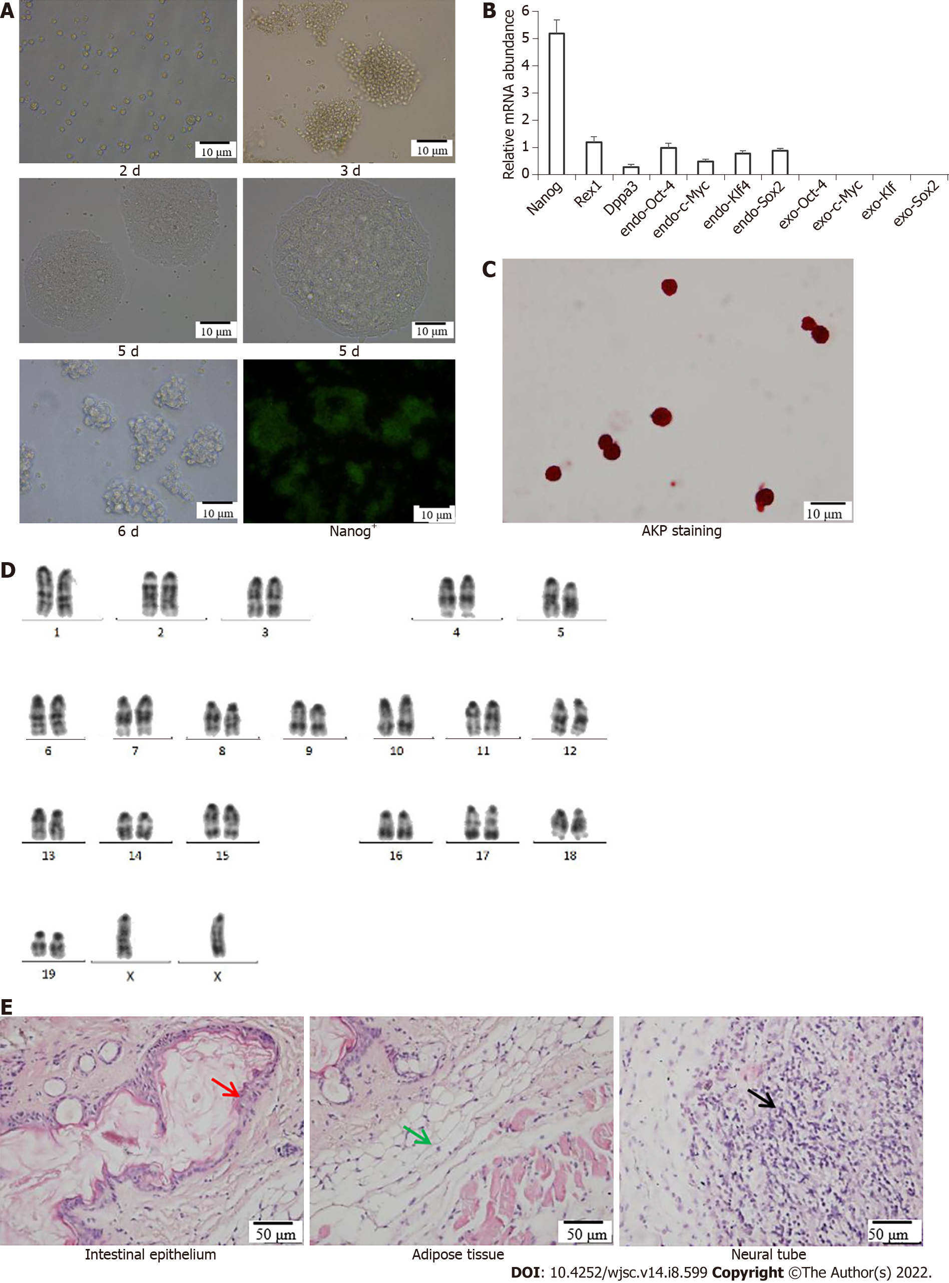
Figure 1 Induced pluripotent stem cells culture and identification.
A: Morphological observation and cell cloning of induced pluripotent stem cells (iPSCs) at 2 d, 3 d, 5 d and 6 d and Nanog+-iPSCs in culture; B: Quantitative polymerase chain reaction identification; C: Alkaline phosphatase staining; D: Chromosome identification; E: Teratoma formation; red arrow = intestinal epithelium (endoderm), green arrow = muscle tissue (mesoderm), and black arrow = nerve tissue (endoderm). AKP: Alkaline phosphatase.
- Citation: Huang XY, Jin ZK, Dou M, Zheng BX, Zhao XR, Feng Q, Feng YM, Duan XL, Tian PX, Xu CX. Sinomenine promotes differentiation of induced pluripotent stem cells into immature dendritic cells with high induction of immune tolerance. World J Stem Cells 2022; 14(8): 599-615
- URL: https://www.wjgnet.com/1948-0210/full/v14/i8/599.htm
- DOI: https://dx.doi.org/10.4252/wjsc.v14.i8.599


