Copyright
©The Author(s) 2022.
World J Stem Cells. Jul 26, 2022; 14(7): 556-576
Published online Jul 26, 2022. doi: 10.4252/wjsc.v14.i7.556
Published online Jul 26, 2022. doi: 10.4252/wjsc.v14.i7.556
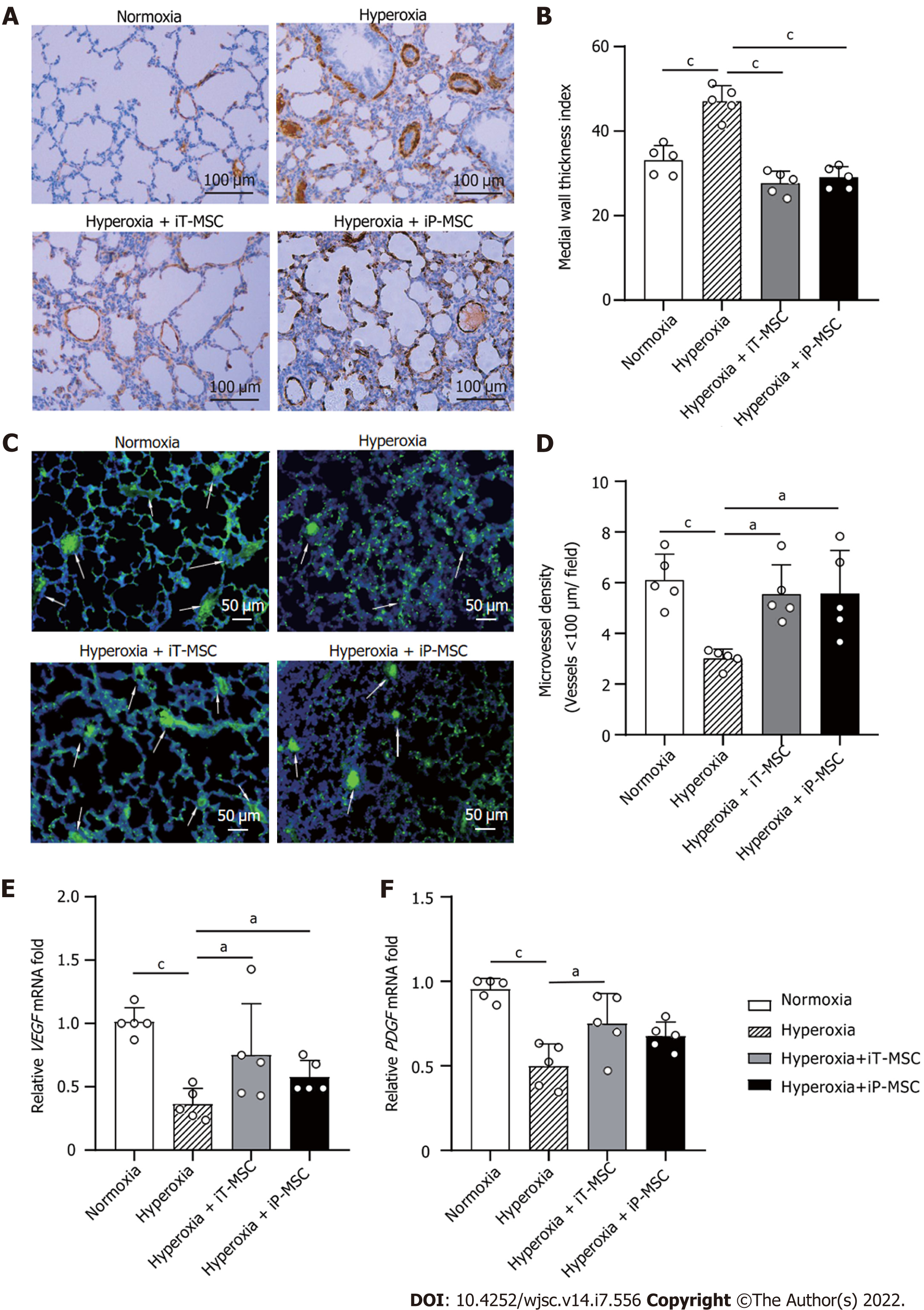
Figure 3 Human umbilical cord-derived mesenchymal stem cells treatment rescues hyperoxia-induced loss of peripheral pulmonary blood vessels and peripheral pulmonary arterial remodeling.
A: Representative photomicrographs of lung sections of rats harvested at postnatal day 21 stained with α smooth muscle actin antibody in normal air or hyperoxia exposure groups, with or without human umbilical cord-derived mesenchymal stem cells administration (scale bars = 100 μm); B and D: Medial thickness index and microvessel density in lungs treated as indicated were calculated to assess hyperoxia-induced peripheral pulmonary vascular remodeling and loss of blood vessels in the peripheral microvasculature (n = 5 for each group, 5 fields/animal); C: Representative lung slides with von Willebrand factor- immunofluorescence staining obtained at 200 × magnification. White arrows highlight stained pulmonary vessels (scale bars = 50 μm); E and F: Vascular endothelial-derived growth factor and platelet-derived growth factor mRNA expression in the lung tissues in the indicated groups. aP < 0.05; bP < 0.01; cP < 0.001. iT: Intratracheal; iP: Intraperitoneal; VEGF: Vascular endothelial-derived growth factor; PDGF: Platelet-derived growth factor; MSC: Mesenchymal stem cell.
- Citation: Dong N, Zhou PP, Li D, Zhu HS, Liu LH, Ma HX, Shi Q, Ju XL. Intratracheal administration of umbilical cord-derived mesenchymal stem cells attenuates hyperoxia-induced multi-organ injury via heme oxygenase-1 and JAK/STAT pathways. World J Stem Cells 2022; 14(7): 556-576
- URL: https://www.wjgnet.com/1948-0210/full/v14/i7/556.htm
- DOI: https://dx.doi.org/10.4252/wjsc.v14.i7.556


