Copyright
©The Author(s) 2022.
World J Stem Cells. Feb 26, 2022; 14(2): 163-182
Published online Feb 26, 2022. doi: 10.4252/wjsc.v14.i2.163
Published online Feb 26, 2022. doi: 10.4252/wjsc.v14.i2.163
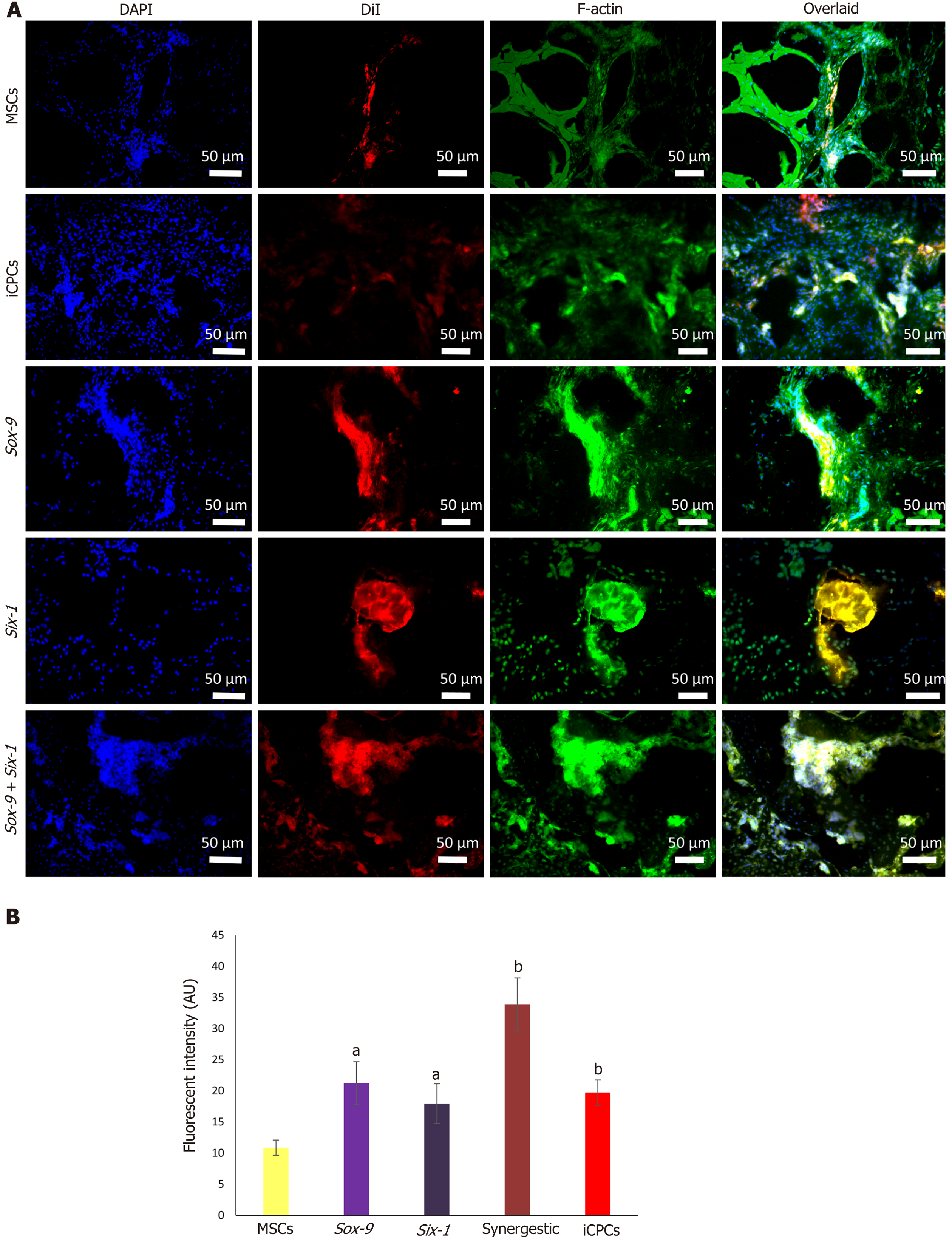
Figure 8 Tracking of transplanted cells in rat intervertebral disc degeneration model.
A: Tracking of the DiI-labeled normal and transfected mesenchymal stem cells (MSCs), and induced chondro-progenitor cells transplanted disc indicated the co-localization of red fluorescence originating from the DiI-labeled cells and green fluorescence from Alexa fluor 488 labeled phalloidin (F-actin), confirming their distribution, and homing in the intervertebral discs; B: Fluorescence intensities of the group transplanted with transfected cells showed significantly high fluorescence compared to normal MSCs. aP < 0.05 vs MSCs; bP < 0.01 vs MSCs. iCPCs: Induced chondro-progenitor cells.
- Citation: Khalid S, Ekram S, Salim A, Chaudhry GR, Khan I. Transcription regulators differentiate mesenchymal stem cells into chondroprogenitors, and their in vivo implantation regenerated the intervertebral disc degeneration. World J Stem Cells 2022; 14(2): 163-182
- URL: https://www.wjgnet.com/1948-0210/full/v14/i2/163.htm
- DOI: https://dx.doi.org/10.4252/wjsc.v14.i2.163


