Copyright
©The Author(s) 2021.
World J Stem Cells. May 26, 2021; 13(5): 452-469
Published online May 26, 2021. doi: 10.4252/wjsc.v13.i5.452
Published online May 26, 2021. doi: 10.4252/wjsc.v13.i5.452
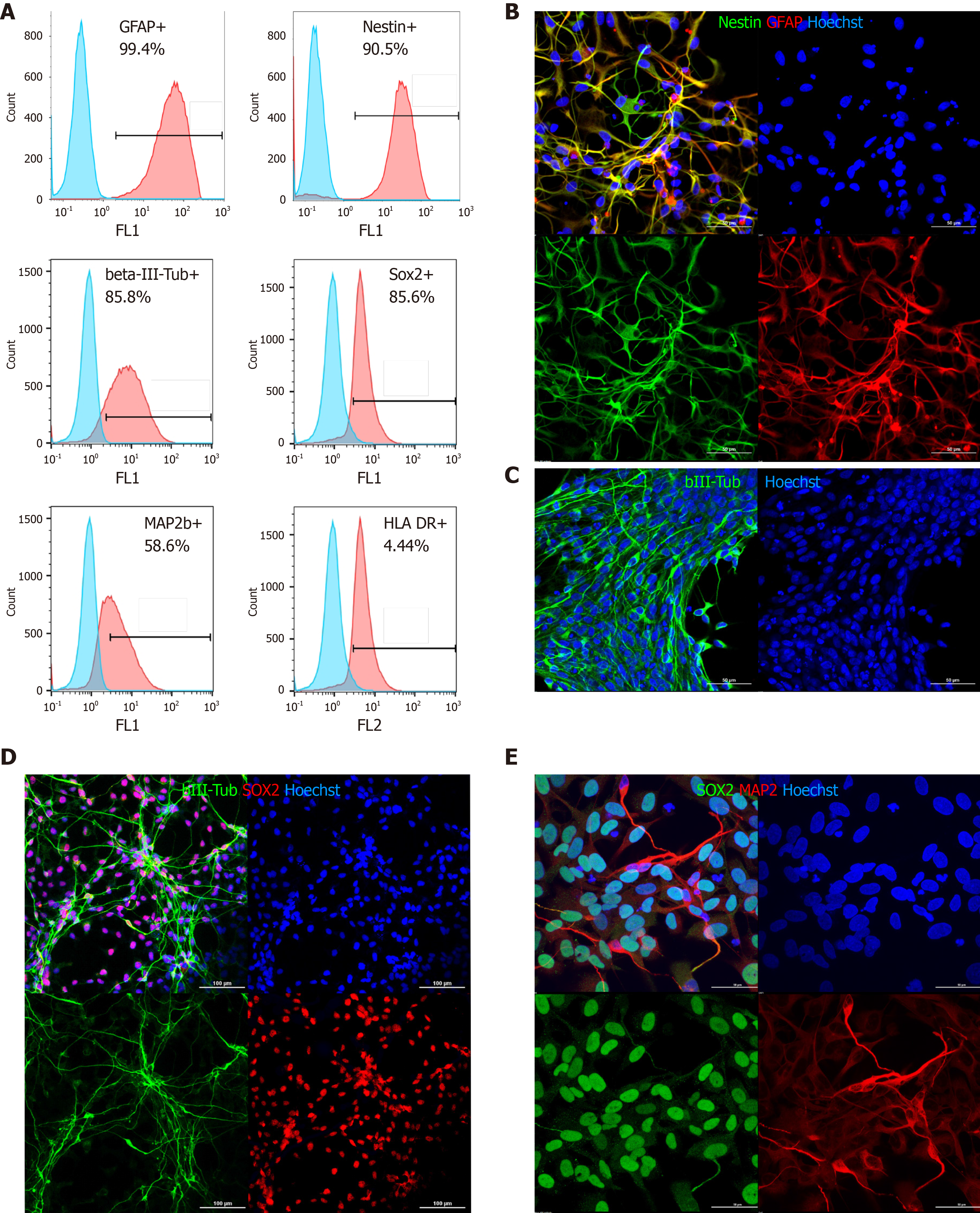
Figure 1 Phenotypic characterization of directly reprogrammed neural precursor cells by flow cytometry and immunocytochemistry.
A: Flow cytometry: blue peaks-negative control (isotype immunoglobulins); from left to right, top to bottom: Glial fibrillary acidic protein, nestin, βIII-tubulin SRY-box transcription factor 2 (SOX2), microtubule associated protein 2 (MAP2), human leukocyte antigen (HLA)-DR; B: Nestin and glial fibrillary acidic protein staining (most cells are double positive); C: βIII-tubulin (green); D: βIII-tubulin (green) and SOX2 (red); E: MAP2 (red) and SOX2 (green) (D and E: Partial spontaneous differentiation of directly reprogrammed neural precursor cells on laminin/poly-L-lysine coated plastic). In all panels, nuclei are counterstained with Hoechst (blue). Scale bar, 50 μm (B, C, and E) and 100 μm (D).
- Citation: Baklaushev VP, Durov OV, Kalsin VA, Gulaev EV, Kim SV, Gubskiy IL, Revkova VA, Samoilova EM, Melnikov PA, Karal-Ogly DD, Orlov SV, Troitskiy AV, Chekhonin VP, Averyanov AV, Ahlfors JE. Disease modifying treatment of spinal cord injury with directly reprogrammed neural precursor cells in non-human primates. World J Stem Cells 2021; 13(5): 452-469
- URL: https://www.wjgnet.com/1948-0210/full/v13/i5/452.htm
- DOI: https://dx.doi.org/10.4252/wjsc.v13.i5.452


