Copyright
©The Author(s) 2021.
World J Stem Cells. May 26, 2021; 13(5): 386-415
Published online May 26, 2021. doi: 10.4252/wjsc.v13.i5.386
Published online May 26, 2021. doi: 10.4252/wjsc.v13.i5.386
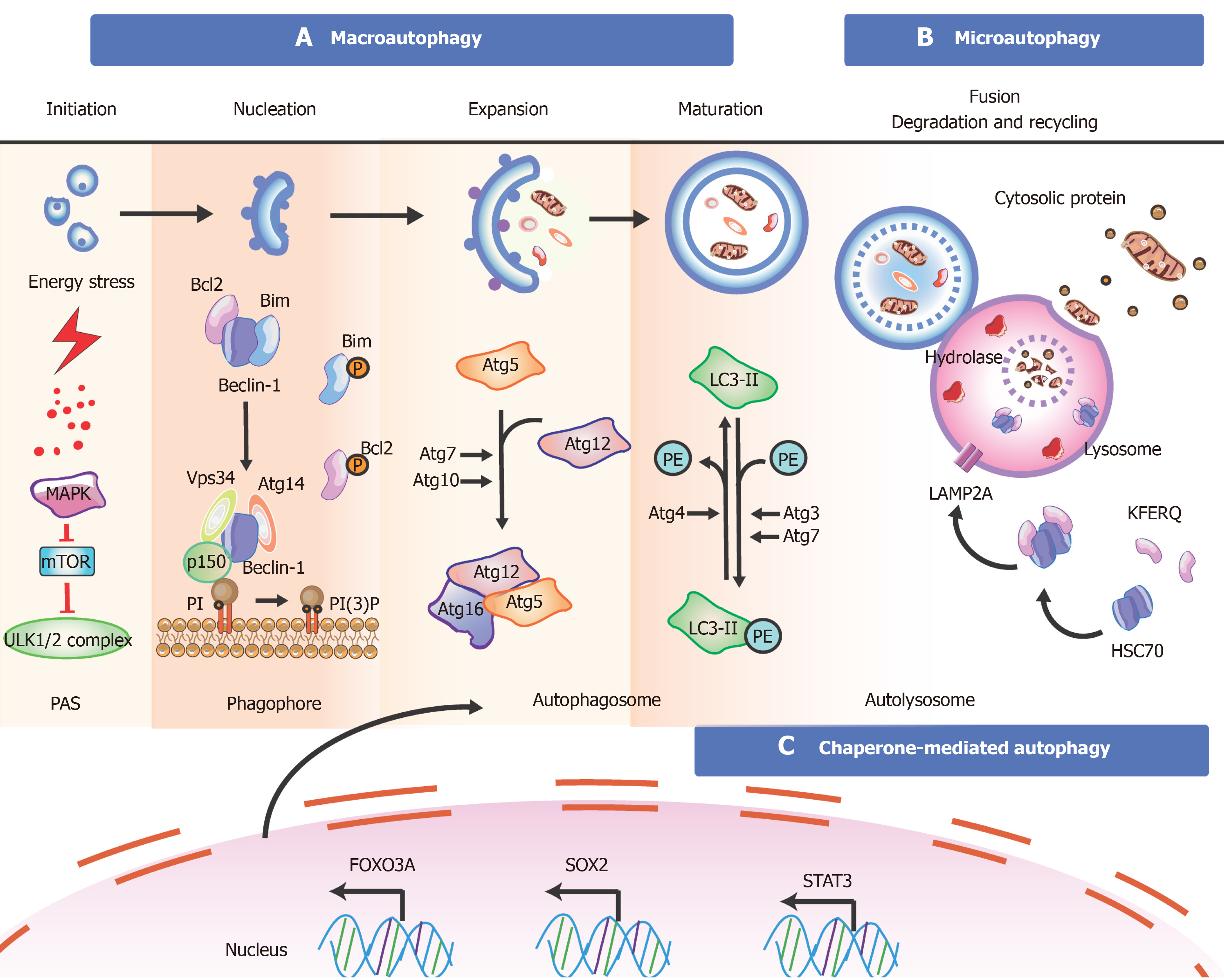
Figure 3 Overview of the mechanisms during autophagy in stem cells.
There are three types of autophagy [macroautophagy (section a), microautophagy (section b), and chaperone-mediated autophagy (section c)] based on different pathways; however, they produce the same results. Besides these proteins, key transcription factors closely related to autophagy are shown. The T-shaped lines indicate inhibitory interactions involved in this pathway, while the solid arrows indicate activating interactions. A: Typically, the mTORC1 complex functions as an inhibitor to control the initiation of autophagy. Under environmental stresses and physiological stressors, AMPK is activated to inhibit the activity of mTORC1, leading to a release of the ULK1 (Unc-51-like kinase complex, also known as ATG1) complex to induce autophagy. This initiation process is known as the phagophore assembly site (PAS) formation. Next, PI3 is phosphorylated to PI3P via the class III PI3-kinase-Beclin1 complex formed by core subunits of Beclin1 (Atg6), Atg14 L, and Vps34-Vps15, resulting in autophagosome formation. The Atg12-Atg5-Atg16L1 complex acts as a regulator for enveloping and translocating the cytoplasmic cargo to the lysosome in misfolded-protein degradation. Atg4 can cleave LC3 (Atg8) to generate cytosolic LC3-I. Atg3 (E2 enzymes) and Atg7 (E1-like enzymes) can lead the conjugation of PE to LC3-I to form lipidated LC3-II, which is combined with the autophagosome membrane to complete and elongate autophagosome formation. Finally, the autophagosome contents undergo degradation due to low lysosomal pH; B: In microautophagy, misfolded or/and toxic proteins can be directly engulfed by the lysosomal membrane and degraded in the lysosome; C: During chaperone-mediated autophagy, the heat shock cognate 70 kDa protein (HSC70) chaperones attach to the pentapeptide motif KFERQ (namely Lys-Phe-Glu-Arg-Gln) for delivery to lysosomes via a specific receptor LAMP2A. Also, some of the key transcription factors are closely linked to the stem cell state and the occurrence of autophagy (bottom). FOXO3A can enhance autophagosome formation via autophagy gene expression in hematopoietic stem cells and breast cancer stem-like cells, which is needed to mitigate an energy crisis and allow cell survival. Besides FOXO3A, other transcription factors such as SOX2, STAT3, OCT4, KLF4, and c-Myc are also vital for reprogramming in the initial creation of stem cells at the genetic level during autophagy.
- Citation: Hu XM, Zhang Q, Zhou RX, Wu YL, Li ZX, Zhang DY, Yang YC, Yang RH, Hu YJ, Xiong K. Programmed cell death in stem cell-based therapy: Mechanisms and clinical applications. World J Stem Cells 2021; 13(5): 386-415
- URL: https://www.wjgnet.com/1948-0210/full/v13/i5/386.htm
- DOI: https://dx.doi.org/10.4252/wjsc.v13.i5.386


