Copyright
©The Author(s) 2021.
World J Stem Cells. Apr 26, 2021; 13(4): 317-330
Published online Apr 26, 2021. doi: 10.4252/wjsc.v13.i4.317
Published online Apr 26, 2021. doi: 10.4252/wjsc.v13.i4.317
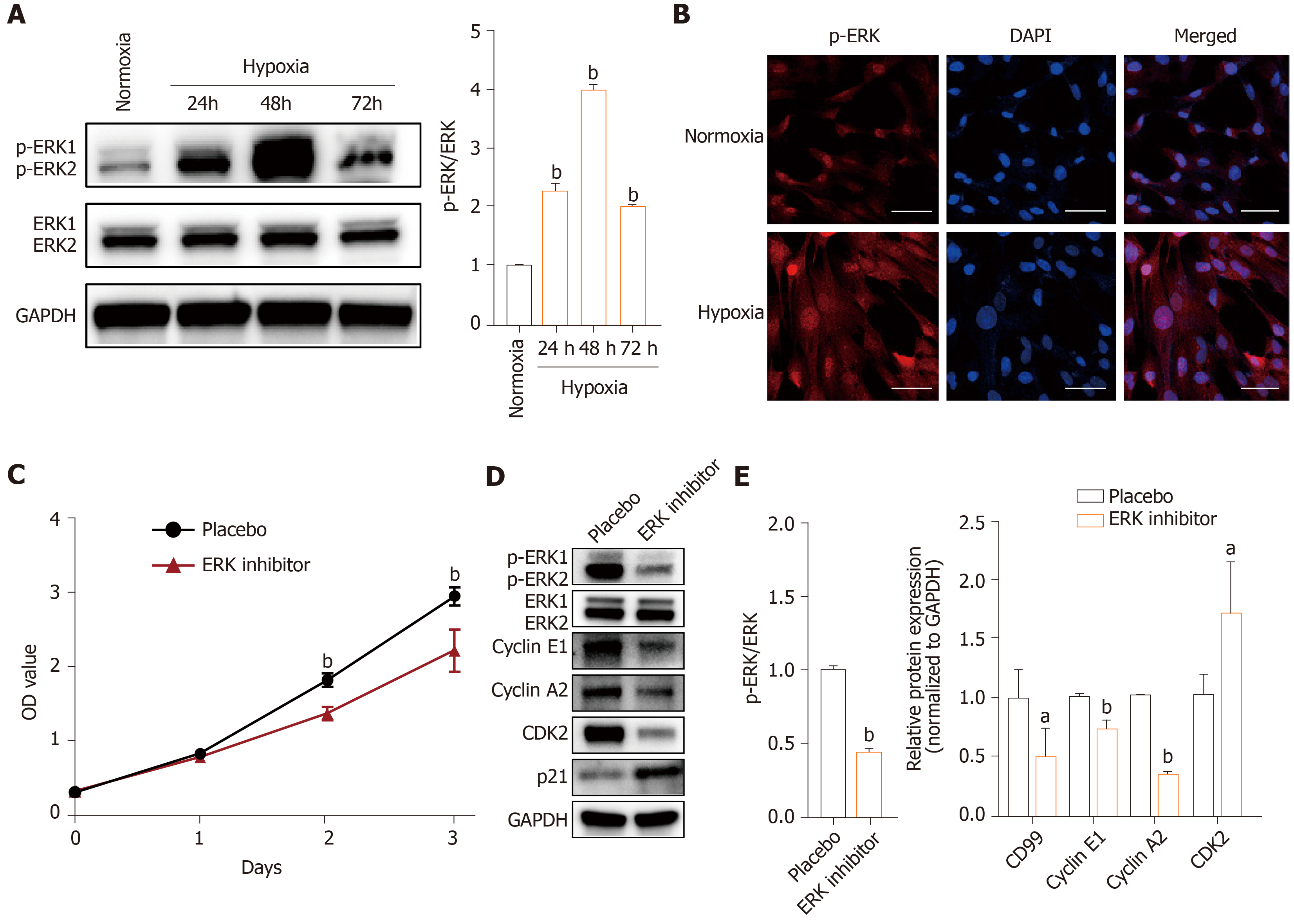
Figure 5 Hypoxia activates MAPK/ERK signaling and promotes human placenta-derived mesenchymal stem cells proliferation.
A: Western blotting of phosphorylated (p)-ERK) in human placenta-derived mesenchymal stem cells (hP-MSCs) exposed to hypoxia for 24, 48, or 72 h normalized against ERK; B: Immunofluorescence microscopy of p-ERK in hP-MSCs cultured under hypoxia and normoxia for 48 h (× 20 magnification; scale bars, 50 μm); C: Proliferation of hP-MSCs cultured under hypoxia after pretreatment with the ERK1/2 signaling inhibitor PD98059 (50 μmol/L). Data are means ± SD (n = 4); D: Western blotting of p-ERK, ERK, cyclin E1, cyclin A2, CDK2, and p21 in hP-MSCs cultured under hypoxia for 48 h after pretreatment with PD98059; E: Expression of p-ERK normalized against that of ERK and cyclin E1, cyclin A2, CDK2, and p21 expression normalized against that of glyceraldehyde-3-phosphate dehydrogenase (GAPDH). Data are means ± SD (n = 3). aP < 0.05 and bP < 0.01. p-ERK: OD: Optical density.
- Citation: Feng XD, Zhu JQ, Zhou JH, Lin FY, Feng B, Shi XW, Pan QL, Yu J, Li LJ, Cao HC. Hypoxia-inducible factor-1α–mediated upregulation of CD99 promotes the proliferation of placental mesenchymal stem cells by regulating ERK1/2. World J Stem Cells 2021; 13(4): 317-330
- URL: https://www.wjgnet.com/1948-0210/full/v13/i4/317.htm
- DOI: https://dx.doi.org/10.4252/wjsc.v13.i4.317


