Copyright
©The Author(s) 2021.
World J Stem Cells. Dec 26, 2021; 13(12): 1905-1917
Published online Dec 26, 2021. doi: 10.4252/wjsc.v13.i12.1905
Published online Dec 26, 2021. doi: 10.4252/wjsc.v13.i12.1905
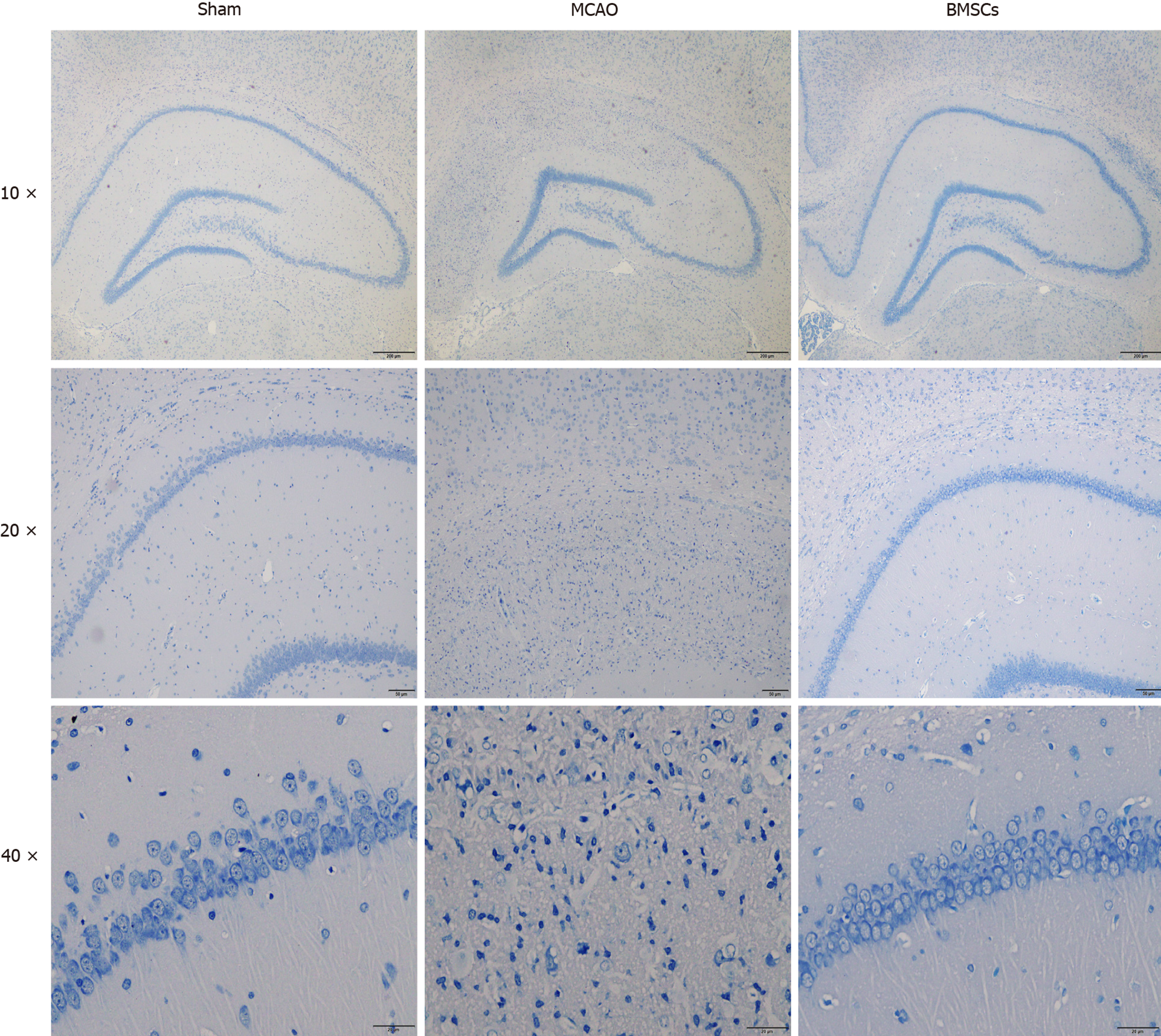
Figure 2 Histopathological changes in brain tissue of rats.
Nissl staining in the hippocampal CA1 region for the Sham, middle cerebral artery occlusion, and bone marrow mesenchymal stem cells group exhibited brain injury after 21 d post-stroke (n = 3). Pathological observation of the hippocampus (magnification, × 40). CA1 region of the hippocampus (magnification, × 100). The morphologies of neurons in the hippocampal CA1 region (magnification, × 400). BMSCs: Bone marrow mesenchymal stem cells; MCAO: Middle cerebral artery occlusion.
- Citation: Zhao LN, Ma SW, Xiao J, Yang LJ, Xu SX, Zhao L. Bone marrow mesenchymal stem cell therapy regulates gut microbiota to improve post-stroke neurological function recovery in rats. World J Stem Cells 2021; 13(12): 1905-1917
- URL: https://www.wjgnet.com/1948-0210/full/v13/i12/1905.htm
- DOI: https://dx.doi.org/10.4252/wjsc.v13.i12.1905


