Copyright
©The Author(s) 2021.
World J Stem Cells. Dec 26, 2021; 13(12): 1905-1917
Published online Dec 26, 2021. doi: 10.4252/wjsc.v13.i12.1905
Published online Dec 26, 2021. doi: 10.4252/wjsc.v13.i12.1905
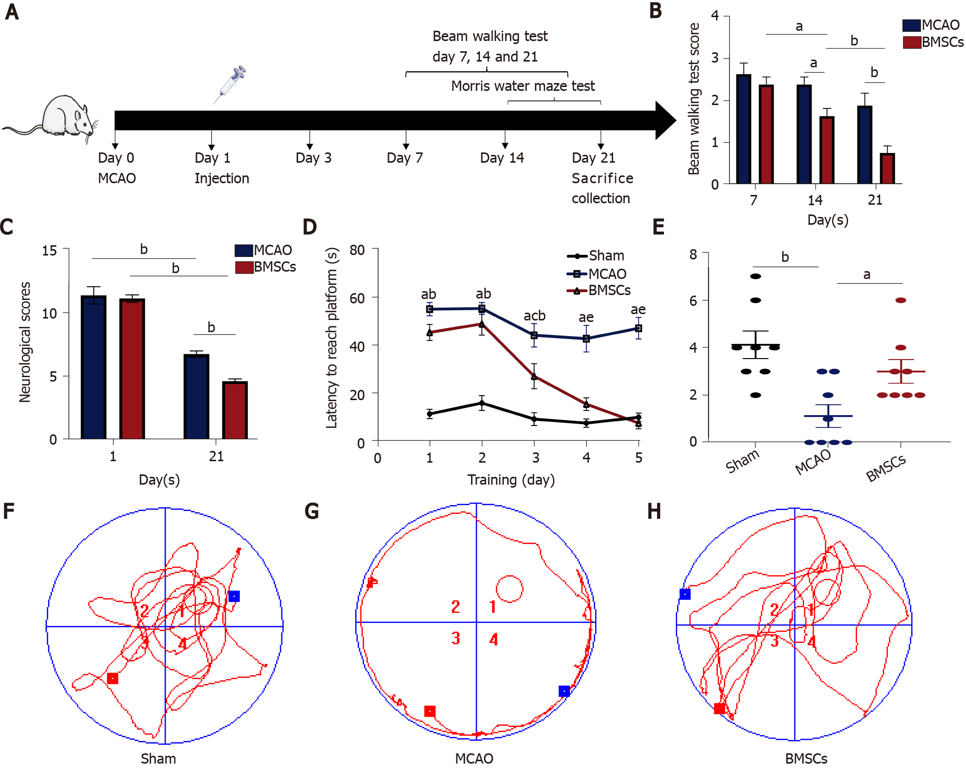
Figure 1 Bone marrow mesenchymal stem cells improve neurological function after stroke.
A: Experimental design. Rats (3-5 wk) were randomly divided into three groups: Sham, middle cerebral artery occlusion (MCAO), and bone marrow mesenchymal stem cells (BMSCs). BMSCs or PBS were injected through the tail vein 1 d after MCAO. The modified Neurological Severity Score (mNSS), the beam walking test, and the Morris water maze test were evaluated before rats were killed after 21 d of reperfusion; B: mNSS was performed at days 1 and 21 after MCAO (aP < 0.05; bP < 0.01); C: Beam walking test were performed at days 7, 14, and 21 after MCAO (bP < 0.01); D-H: Morris water maze test. The time that rats needed to escape latency to find the hidden platform (D). aP < 0.01 when Sham vs MCAO; bP < 0.01 when Sham vs BMSCs; cP < 0.01 when Sham vs BMSCs; dP < 0.01 when MCAO vs BMSCs; eP < 0.01 when MCAO vs BMSCs. The number of rats crossing over the target platform on the sixth day (aP < 0.05; bP < 0.01) (E). The data are expressed as the mean ± SEM (n = 10). The tracks of each group on the sixth day (F-H). BMSCs: Bone marrow mesenchymal stem cells; MCAO: Middle cerebral artery occlusion.
- Citation: Zhao LN, Ma SW, Xiao J, Yang LJ, Xu SX, Zhao L. Bone marrow mesenchymal stem cell therapy regulates gut microbiota to improve post-stroke neurological function recovery in rats. World J Stem Cells 2021; 13(12): 1905-1917
- URL: https://www.wjgnet.com/1948-0210/full/v13/i12/1905.htm
- DOI: https://dx.doi.org/10.4252/wjsc.v13.i12.1905


