Copyright
©The Author(s) 2021.
World J Stem Cells. Nov 26, 2021; 13(11): 1783-1796
Published online Nov 26, 2021. doi: 10.4252/wjsc.v13.i11.1783
Published online Nov 26, 2021. doi: 10.4252/wjsc.v13.i11.1783
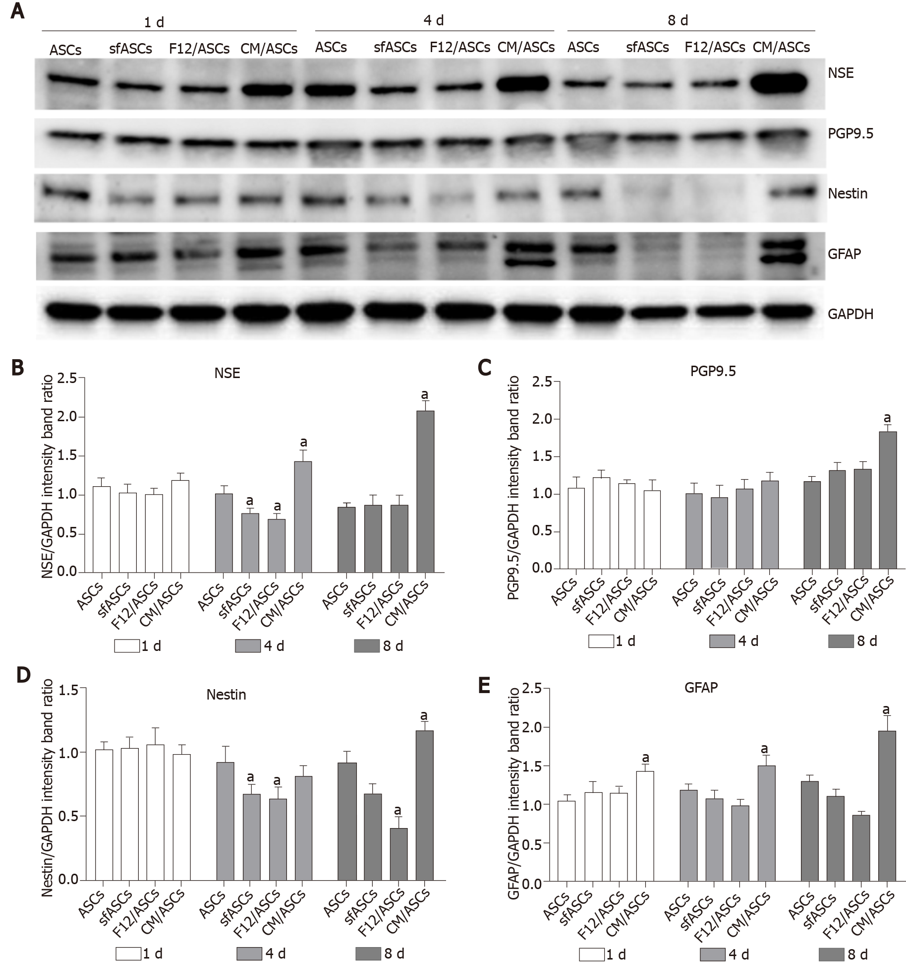
Figure 3 Western blot analysis of neural marker expression in different samples of adipose-derived stem cells cultures.
A: Immunoblot analysis of whole-cell lysates at day 1, 4 and 8 of culture for NSE, PGP9.5, nestin, GFAP and GAPDH as internal control; B-E: ASCs: Control adipose-derived stem cells (ASCs) cultured in basal Dulbecco's Modified Eagle Medium (DMEM); sfASCs: ASCs cultured in serum-free DMEM; F12/ASCs: ASCs cultured in serum-free DMEM/F12; CM/ASCs: ASCs cultured in serum-free DMEM/F12 conditioned from ARPE-19. Quantitative data are illustrated in histograms. Values are expressed as mean ± SD obtained from three independent experiments. aP < 0.05 vs ASCs of corresponding time point; Two-way ANOVA, followed by Tukey’s multiple comparisons test. NSE: Neuron specific enolase; PGP9.5: Protein gene product 9.5; GFAP: Glial fibrillary acidic protein; GAPDH: Glyceraldehyde 3-phosphate dehydrogenase; sfASCs: Serum-free adipose-derived stem cells; CM: Conditioned medium.
- Citation: Mannino G, Cristaldi M, Giurdanella G, Perrotta RE, Lo Furno D, Giuffrida R, Rusciano D. ARPE-19 conditioned medium promotes neural differentiation of adipose-derived mesenchymal stem cells. World J Stem Cells 2021; 13(11): 1783-1796
- URL: https://www.wjgnet.com/1948-0210/full/v13/i11/1783.htm
- DOI: https://dx.doi.org/10.4252/wjsc.v13.i11.1783


