Copyright
©The Author(s) 2021.
World J Stem Cells. Nov 26, 2021; 13(11): 1667-1695
Published online Nov 26, 2021. doi: 10.4252/wjsc.v13.i11.1667
Published online Nov 26, 2021. doi: 10.4252/wjsc.v13.i11.1667
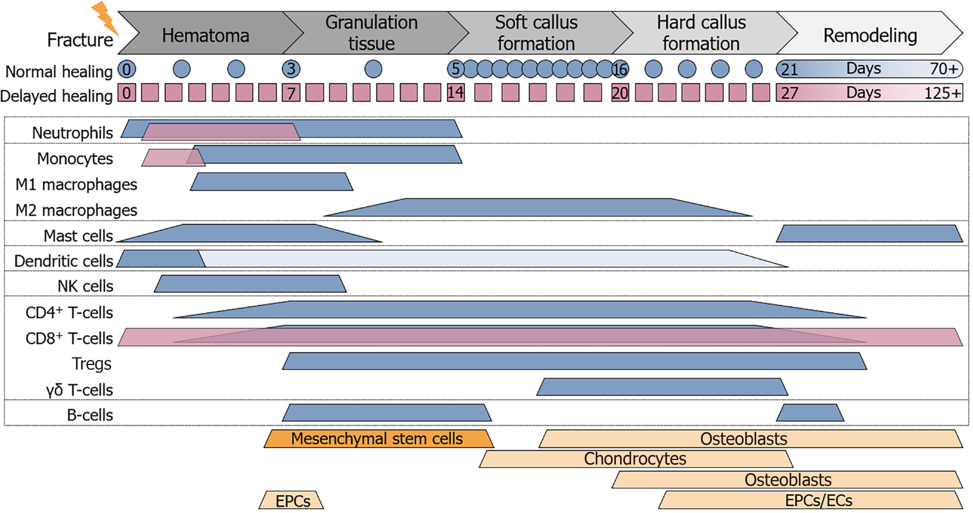
Figure 1 Cell composition at the site of fracture.
During the different phases of fracture healing the cell composition at the site of fracture changes. Expected timeline of normal (blue) and delayed (magenta) fracture healing is depicted below the phases of fracture healing. Colored (blue and magenta) beams representing the timeframe where immune cells are expected to be active at the site of fracture, based on different in vivo studies. Orange beams representing the timeframe where mesenchymal stem cells, chondrocytes, osteoblasts, osteoclasts, endothelial progenitor cells, and endothelial cells are involved in the fracture healing process. CD: Cluster of differentiation; EPCs: Endothelial progenitor cells; ECs: Endothelial cells; NK: Natural killer.
- Citation: Ehnert S, Relja B, Schmidt-Bleek K, Fischer V, Ignatius A, Linnemann C, Rinderknecht H, Huber-Lang M, Kalbitz M, Histing T, Nussler AK. Effects of immune cells on mesenchymal stem cells during fracture healing. World J Stem Cells 2021; 13(11): 1667-1695
- URL: https://www.wjgnet.com/1948-0210/full/v13/i11/1667.htm
- DOI: https://dx.doi.org/10.4252/wjsc.v13.i11.1667


