Copyright
©The Author(s) 2021.
World J Stem Cells. Oct 26, 2021; 13(10): 1595-1609
Published online Oct 26, 2021. doi: 10.4252/wjsc.v13.i10.1595
Published online Oct 26, 2021. doi: 10.4252/wjsc.v13.i10.1595
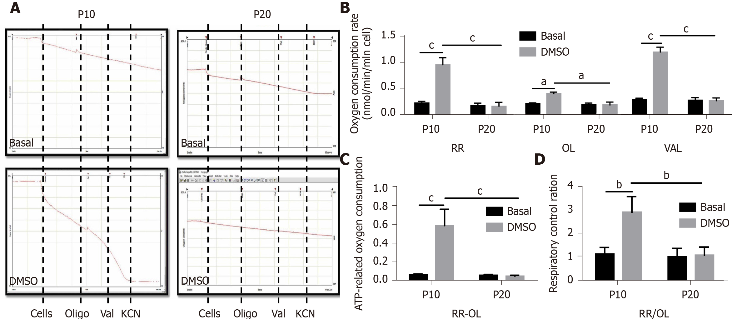
Figure 6 Measurement of mitochondrial respiration in HepaRG cells at passage 10 or passage 20 in basal conditions or after the transdifferentiation protocol (dimethyl sulfoxide).
A: Representative oxymetric traces of mitochondrial respiration in HepaRG cells. Where indicated, the following were added: 4 × 10⁶ HepaRG cells, 8 μg/mL oligomycin (OL), 2 μg/mL valinomycin (VAL), 3 mmol/L potassium cyanide (KCN). The continuous line represents the oxygen concentration measured every 0.1 s throughout the time-course of the assay; B: Representative graph of the normalized and KCN-insensitive-corrected oxygen consumption rates measured under resting respiration (RR) conditions, in the presence of OL and in the presence of VAL (see panel A); C: ATP-dependent oxygen consumption measured as absolute difference between that obtained in the absence and that in the presence of oligomycin (RR-OL); D: Respiratory control ratios obtained dividing the oxygen consumption rates measured under resting conditions by that in the presence of oligomycin (RR/OL). Data in the graphs are represented as the mean ± standard deviation of three independent experiments. Statistical differences were assessed by two-way analysis of variance followed by the Tukey’s test as the post hoc test. aP < 0.05; bP < 0.01; cP < 0.001. DMSO: Dimethyl sulfoxide; P10: Passage 10; P20: Passage 20.
- Citation: Bellanti F, di Bello G, Tamborra R, Amatruda M, Lo Buglio A, Dobrakowski M, Kasperczyk A, Kasperczyk S, Serviddio G, Vendemiale G. Impact of senescence on the transdifferentiation process of human hepatic progenitor-like cells. World J Stem Cells 2021; 13(10): 1595-1609
- URL: https://www.wjgnet.com/1948-0210/full/v13/i10/1595.htm
- DOI: https://dx.doi.org/10.4252/wjsc.v13.i10.1595


