Copyright
©The Author(s) 2021.
World J Stem Cells. Oct 26, 2021; 13(10): 1580-1594
Published online Oct 26, 2021. doi: 10.4252/wjsc.v13.i10.1580
Published online Oct 26, 2021. doi: 10.4252/wjsc.v13.i10.1580
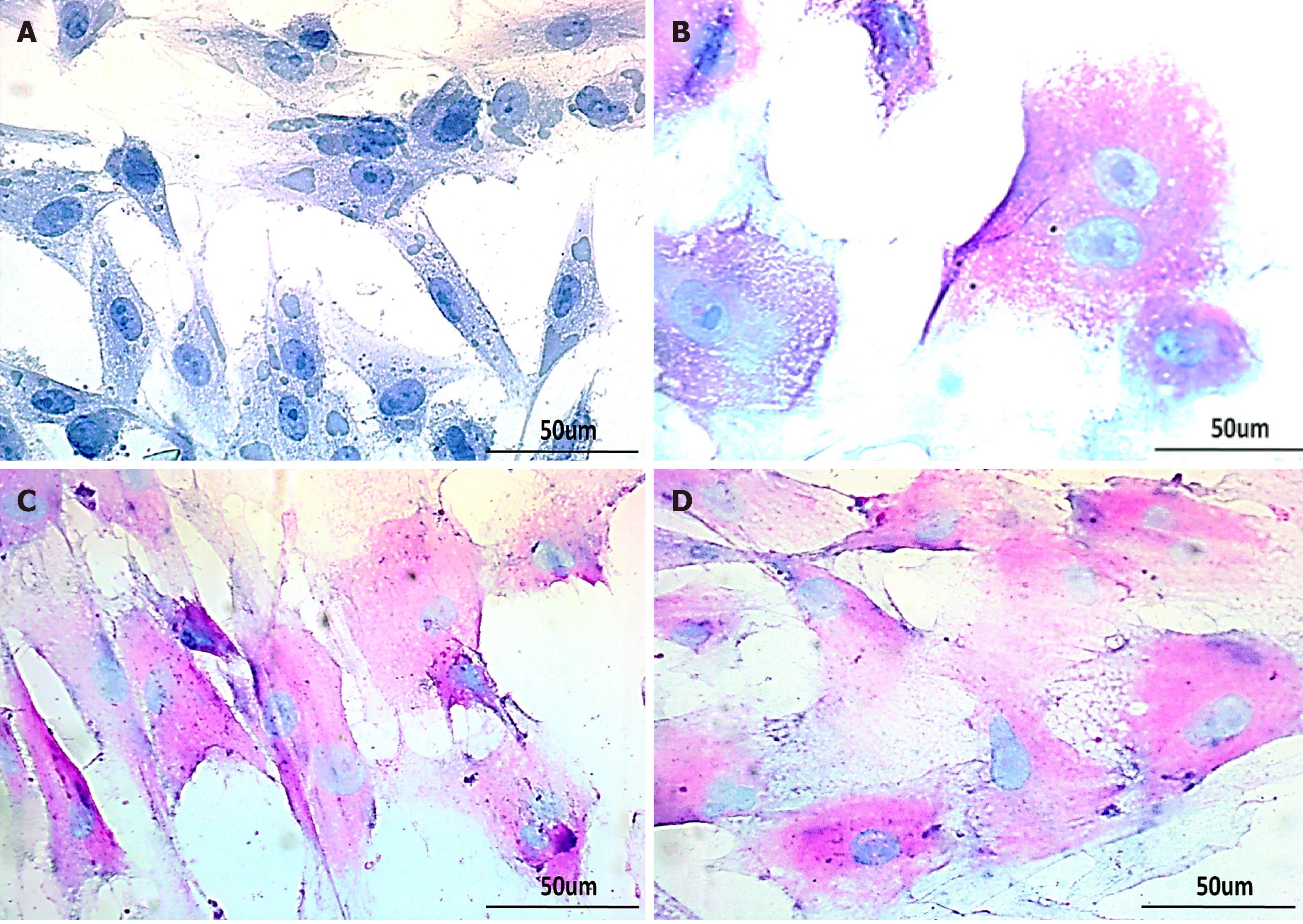
Figure 7 Glycogen detection in treated human umbilical cord-mesenchymal stem cells by periodic acid Schiff staining.
Bright-field images of A: untreated control showing negative periodic acid Schiff staining (PAS); B: MSCs treated with glycyrrhizic acid (GA); C: 18β-glycyrrhetinic acid (GT); and D: GA+GT (combination group) at day 21 showing PAS positive cells stained as purple-pink, indicating the presence of glycogen in their cytoplasm. Bi-nucleated cells (a hepatocyte characteristic) were also observed. Images were taken at 40× magnification.
- Citation: Fatima A, Malick TS, Khan I, Ishaque A, Salim A. Effect of glycyrrhizic acid and 18β-glycyrrhetinic acid on the differentiation of human umbilical cord-mesenchymal stem cells into hepatocytes. World J Stem Cells 2021; 13(10): 1580-1594
- URL: https://www.wjgnet.com/1948-0210/full/v13/i10/1580.htm
- DOI: https://dx.doi.org/10.4252/wjsc.v13.i10.1580


