Copyright
©The Author(s) 2021.
World J Stem Cells. Oct 26, 2021; 13(10): 1446-1479
Published online Oct 26, 2021. doi: 10.4252/wjsc.v13.i10.1446
Published online Oct 26, 2021. doi: 10.4252/wjsc.v13.i10.1446
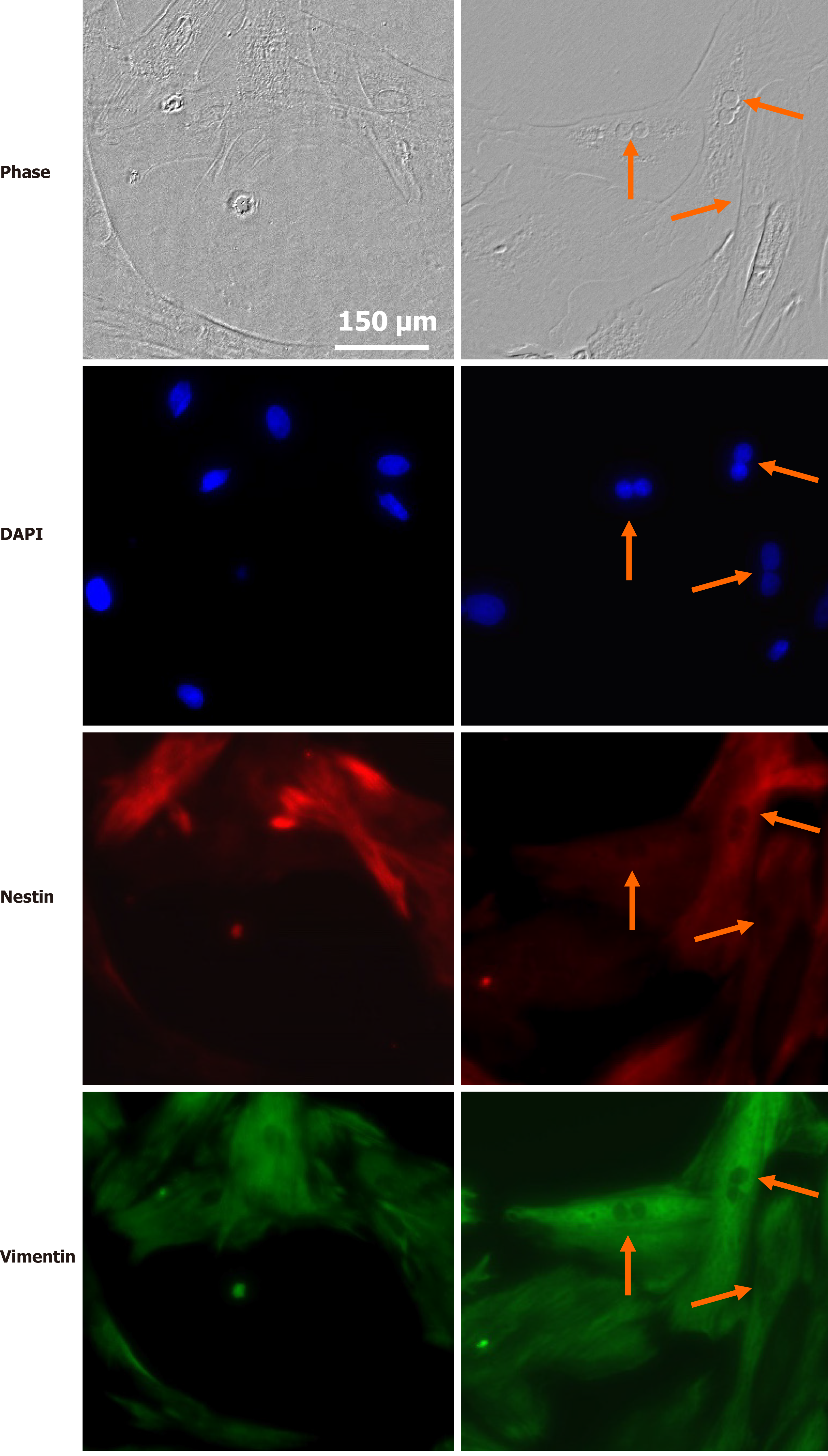
Figure 4 Stem cell properties of Müller glial cells.
Photomicrographs of Müller glial cell cultures from rat retina in interphase (left) or mitosis (self-renewal) (right), showing nuclei labeled with the DNA probe DAPI, expression of the stem cell marker nestin (red) and of vimentin (green). Arrows indicate mitotic anaphases. The bar indicates 150 µm.
- Citation: German OL, Vallese-Maurizi H, Soto TB, Rotstein NP, Politi LE. Retina stem cells, hopes and obstacles. World J Stem Cells 2021; 13(10): 1446-1479
- URL: https://www.wjgnet.com/1948-0210/full/v13/i10/1446.htm
- DOI: https://dx.doi.org/10.4252/wjsc.v13.i10.1446


