Copyright
©The Author(s) 2021.
World J Stem Cells. Jan 26, 2021; 13(1): 91-114
Published online Jan 26, 2021. doi: 10.4252/wjsc.v13.i1.91
Published online Jan 26, 2021. doi: 10.4252/wjsc.v13.i1.91
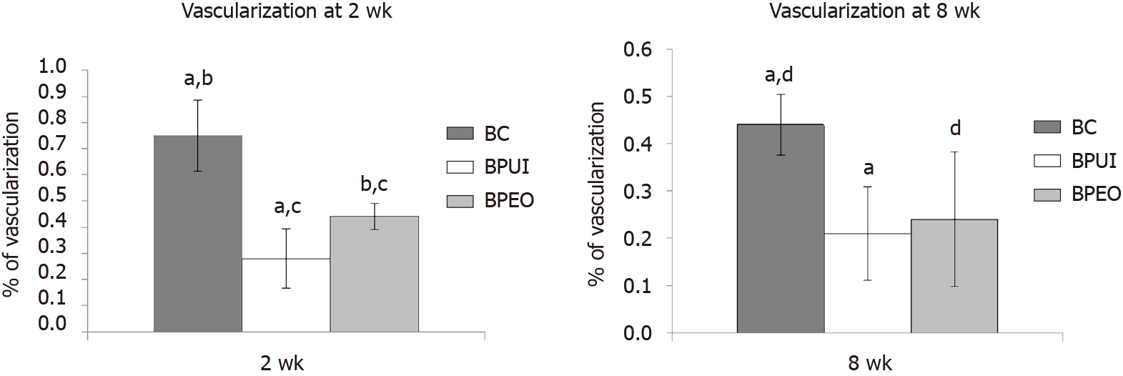
Figure 5 Histomorphometric analysis of ectopic osteogenic implants.
Percentage of vascularization was determined in: A: Two-week-old and B: Eight-week-old BC [implants containing only bone mineral matrix (BMM) carrier], BPUI (implants containing uninduced adipose-derived stem cells, platelet-rich plasma and BMM carrier) and BPEO (implants containing simultaneously applied endothelial and osteogenic differentiated adipose-derived stem cells, platelet-rich plasma and BMM carrier). Four samples (n = 4) per each group per experimental period [n (BC) = 4, n (BPUI) = 4, n (BPEO) = 4] were taken for this analysis. For each group for both experimental periods, results were presented as mean values ± standard deviation. Error bars represent standard deviation. aP < 0.01 BC vs BPUI. bP < 0.01 BC vs BPEO. cP < 0.05 BPUI vs BPEO. dP < 0.05 BC and BPEO. BC: Implants containing only bone mineral matrix carrier; BPUI: Implants containing uninduced adipose-derived stem cells, platelet-rich plasma and bone mineral matrix carrier; BPEO: Implants containing simultaneously applied endothelial and osteogenic differentiated adipose-derived stem cells, platelet-rich plasma and bone mineral matrix carrier.
- Citation: Najdanović JG, Cvetković VJ, Stojanović ST, Vukelić-Nikolić MĐ, Živković JM, Najman SJ. Vascularization and osteogenesis in ectopically implanted bone tissue-engineered constructs with endothelial and osteogenic differentiated adipose-derived stem cells. World J Stem Cells 2021; 13(1): 91-114
- URL: https://www.wjgnet.com/1948-0210/full/v13/i1/91.htm
- DOI: https://dx.doi.org/10.4252/wjsc.v13.i1.91


