Copyright
©The Author(s) 2021.
World J Stem Cells. Jan 26, 2021; 13(1): 128-138
Published online Jan 26, 2021. doi: 10.4252/wjsc.v13.i1.128
Published online Jan 26, 2021. doi: 10.4252/wjsc.v13.i1.128
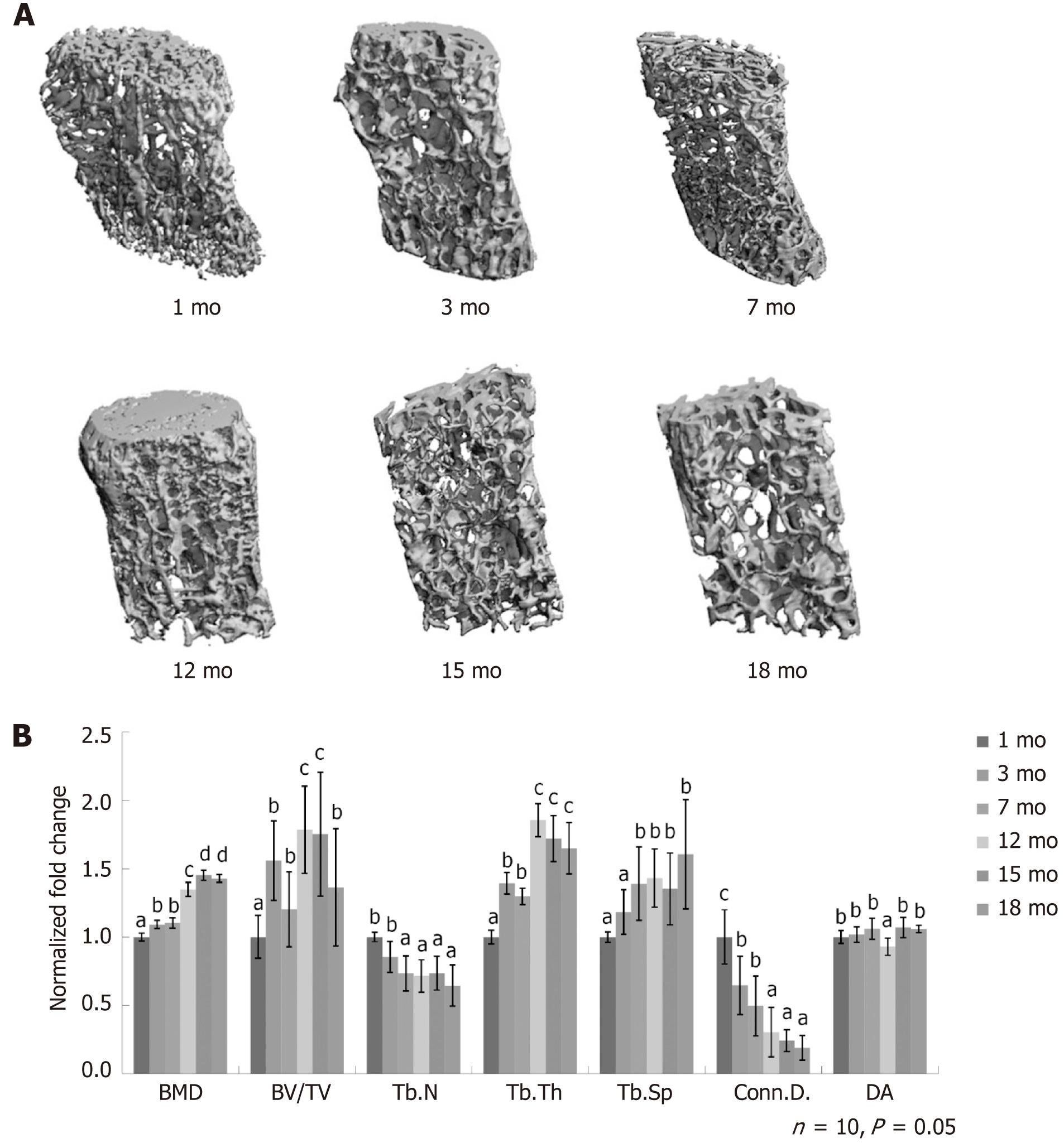
Figure 1 Quantitative measurement of skeletal features.
A: Micro-computed tomography images of L4 lumbar spine at 1 mo, 3 mo, 7 mo, 12 mo, 15 mo, and 18 mo; B: Quantitative measurement of the densitometry and structural parameters of cancellous bone, including the ratio of bone volume to tissue volume (BV/TV), the connectivity density of trabeculae (Conn.D.), the trabecular number (Tb.N), the trabecular thickness (Tb.Th), and the trabecular spaces (Tb.Sp). Bone mass density, Tb.Th, and Tb.Sp increased with age while Tb.N and Conn.D. decreased constantly. The BV/TV increased and reached the plateau at the age of 12 mo. The degree of anisotropy did not change over time between the window of 1 mo and 18 mo. BMD: Bone mass density; BV/TV: Bone volume to tissue volume; Tb.N: Trabecular number; Tb.Th: Trabecular thickness; Tb.Sp: Trabecular spaces; Conn.D.: Connectivity density of trabeculae; DA: Degree of anisotropy.
- Citation: Cheng YH, Liu SF, Dong JC, Bian Q. Transcriptomic alterations underline aging of osteogenic bone marrow stromal cells. World J Stem Cells 2021; 13(1): 128-138
- URL: https://www.wjgnet.com/1948-0210/full/v13/i1/128.htm
- DOI: https://dx.doi.org/10.4252/wjsc.v13.i1.128


