Copyright
©The Author(s) 2020.
World J Stem Cells. Jul 26, 2020; 12(7): 633-658
Published online Jul 26, 2020. doi: 10.4252/wjsc.v12.i7.633
Published online Jul 26, 2020. doi: 10.4252/wjsc.v12.i7.633
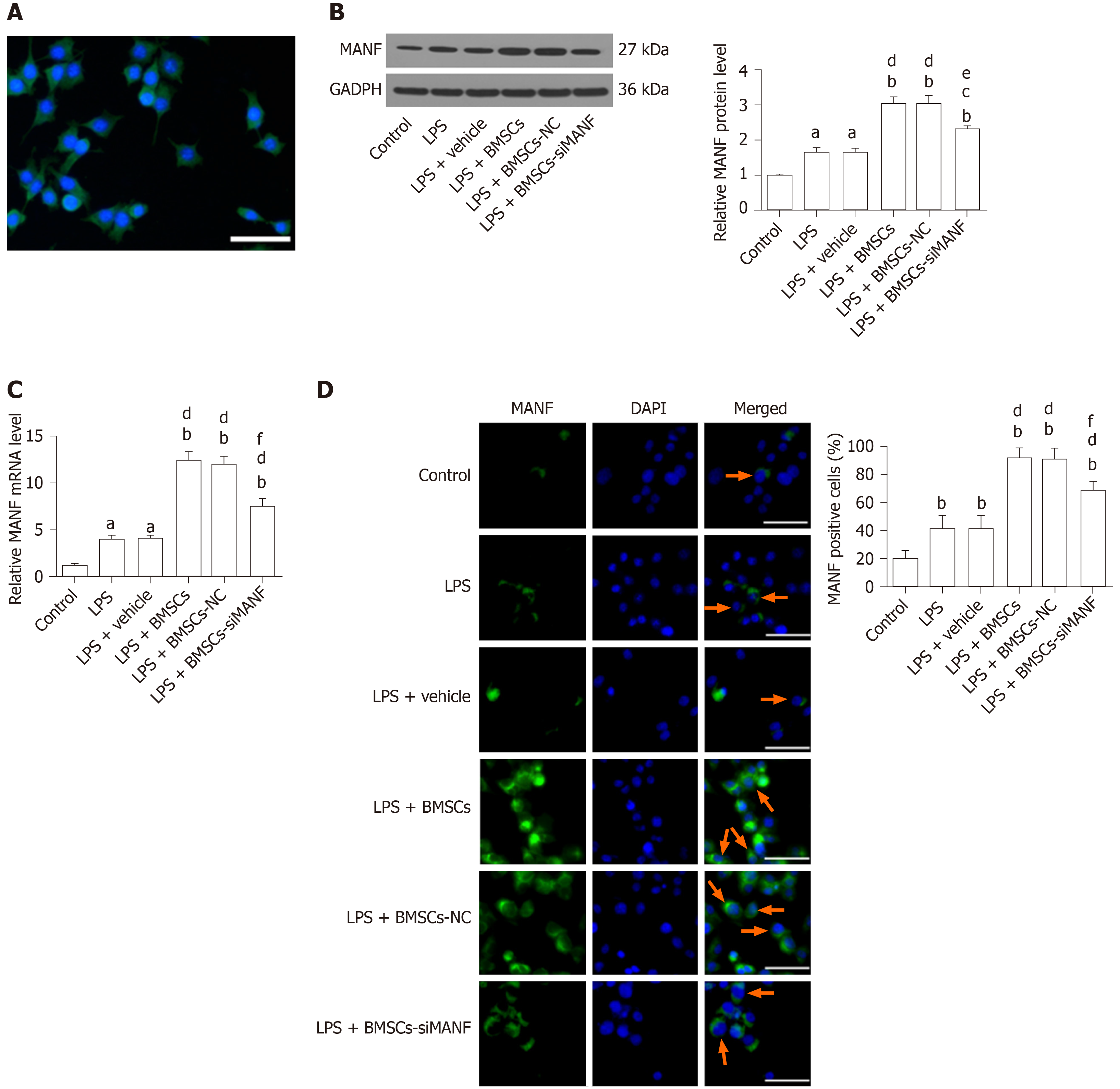
Figure 5 Mesencephalic astrocyte–derived neurotrophic factor expression in lipopolysaccharide-stimulated microglia.
A: Representative immunocytochemical staining for Iba1 (green) and co-staining for DAPI (blue) as a nuclear counterstain. Scale bar = 50 μm; B–D: Western blot (B), qRT-PCR (C), and immunocytochemistry analysis (D) of MANF expression in microglia pretreated with 100 ng/mL LPS for 24 h with or without BMSCs. Representative images of MANF (green), with DAPI (blue) as a nuclear counterstain, are shown. aP < 0.05, bP < 0.01 vs Control group; cP < 0.05, dP < 0.01 vs LPS and LPS + vehicle groups; eP < 0.05, fP < 0.01 vs LPS + BMSCs and LPS + BMSCs-NC groups. Scale bar = 50 μm. Arrows point to the MANF+ cells. The values are expressed as the mean ± SD (n = 6). MANF: Mesencephalic astrocyte–derived neurotrophic factor; GAPDH: Glyceraldehyde-3-phosphate dehydrogenase; LPS: Lipopolysaccharide; BMSCs: Bone marrow mesenchymal stem cells; BMSCs-NC: Negative control-transfected BMSCs; BMSCs-siMANF: MANF siRNA-transfected BMSCs; DAPI: 4'6-diamidino-2-phenylindole.
- Citation: Yang F, Li WB, Qu YW, Gao JX, Tang YS, Wang DJ, Pan YJ. Bone marrow mesenchymal stem cells induce M2 microglia polarization through PDGF-AA/MANF signaling. World J Stem Cells 2020; 12(7): 633-658
- URL: https://www.wjgnet.com/1948-0210/full/v12/i7/633.htm
- DOI: https://dx.doi.org/10.4252/wjsc.v12.i7.633


