Copyright
©The Author(s) 2020.
World J Stem Cells. Jul 26, 2020; 12(7): 633-658
Published online Jul 26, 2020. doi: 10.4252/wjsc.v12.i7.633
Published online Jul 26, 2020. doi: 10.4252/wjsc.v12.i7.633
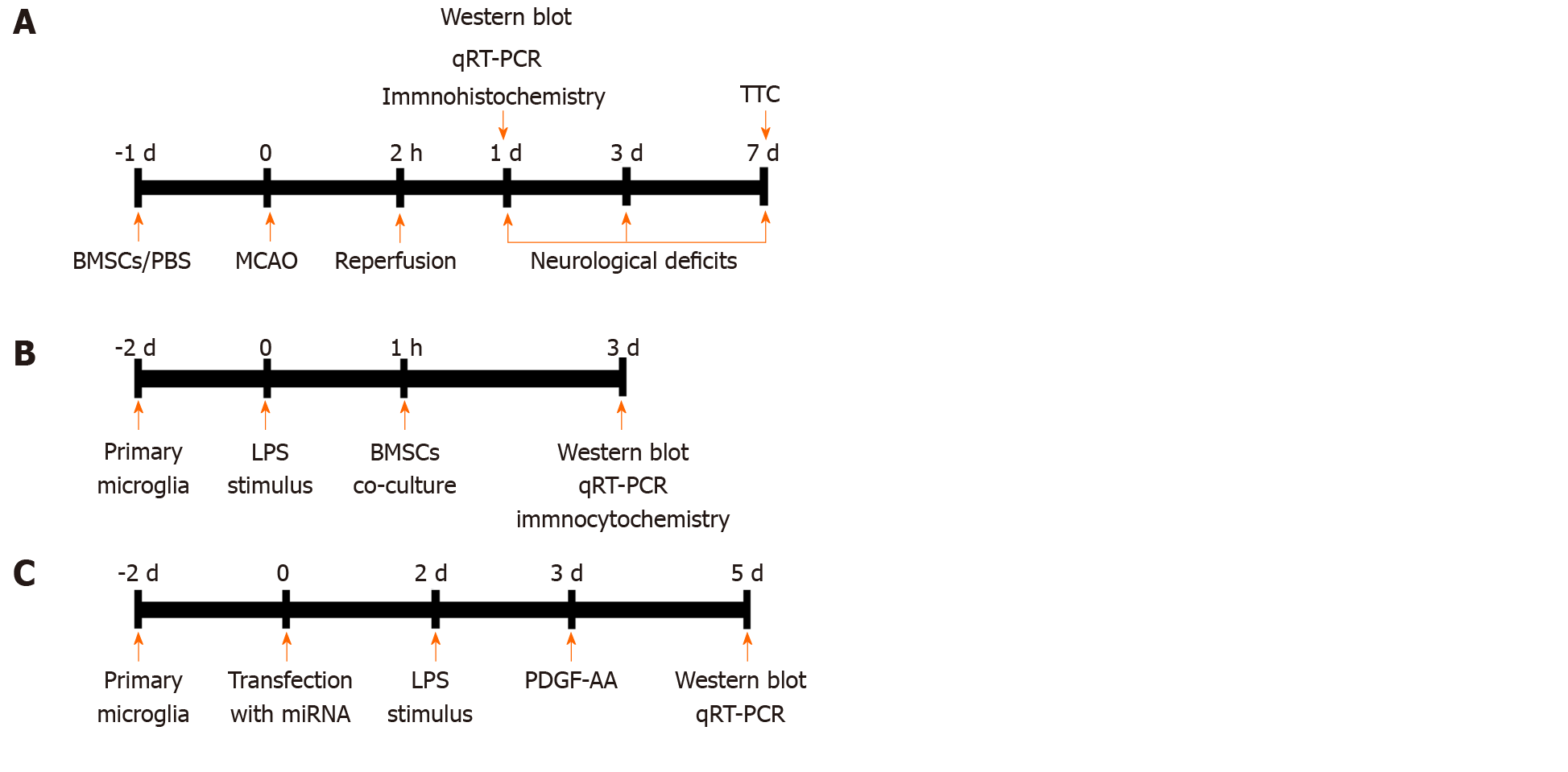
Figure 1 Experimental design.
A: In vivo experiments. Bone marrow mesenchymal stem cells (BMSCs) or PBS were infused into the right striatum 1 d before MCAO. Animals were killed at 1 d for analysis by Western blot, qRT-PCR, and immunohistochemistry. Neurological function tests were evaluated at 1, 3, and 7 d post-stroke. Rats were killed at 7 d for TTC staining and measurement of infarct volume. B and C: In vitro experiments. Primary microglial cells were cultured for 2 d and exposed to 100 ng/mL LPS followed by indirect (Transwell) BMSCs coculture. After 2 d, cocultures were analyzed by Western blot, qRT-PCR, and immunocytochemistry (B). Cultured HAPI cells were transfected with miR-mimics, anti-miR, or corresponding negative control oligonucleotides for 24 h. PDGF-AA was added to the culture medium of HAPI cells pretreated with 100 ng/mL LPS for 24 h. After 2 d, cells were collected for Western blot and qRT-PCR (C). BMSCs: Bone marrow mesenchymal stem cells; PBS: Phosphate-buffered saline; MCAO: Middle cerebral artery occlusion; qRT-PCR: Real-time quantitative reverse transcription-polymerase chain reaction; TTC: 2,3,5-triphenylterazolium chloride; LPS: Lipopolysaccharide; PDGF-AA: Platelet-derived growth factor-AA.
- Citation: Yang F, Li WB, Qu YW, Gao JX, Tang YS, Wang DJ, Pan YJ. Bone marrow mesenchymal stem cells induce M2 microglia polarization through PDGF-AA/MANF signaling. World J Stem Cells 2020; 12(7): 633-658
- URL: https://www.wjgnet.com/1948-0210/full/v12/i7/633.htm
- DOI: https://dx.doi.org/10.4252/wjsc.v12.i7.633


