Copyright
©The Author(s) 2020.
World J Stem Cells. Dec 26, 2020; 12(12): 1652-1666
Published online Dec 26, 2020. doi: 10.4252/wjsc.v12.i12.1652
Published online Dec 26, 2020. doi: 10.4252/wjsc.v12.i12.1652
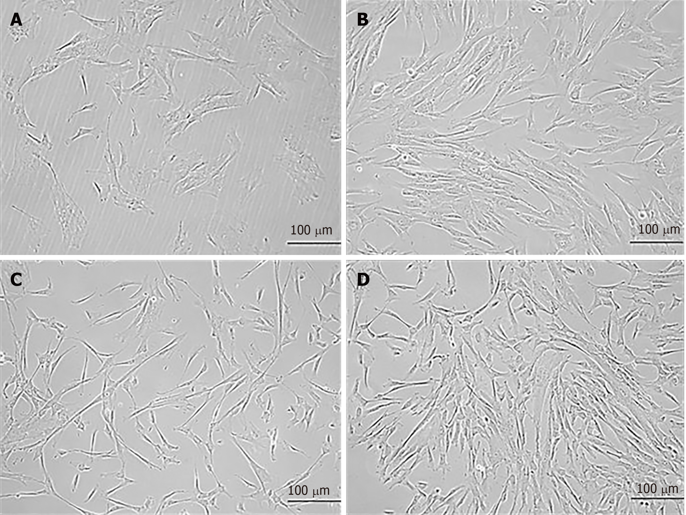
Figure 1 Morphology of mesenchymal stem cells at different passages.
Phase contrast images (20 ×) of human umbilical cord-derived mesenchymal stem cells show fibroblast-like morphology at passage 0 (P0) at day 10 and day 12 of culture. Similarly, cells at passage 1 (P1) show fibroblast-like morphology with extensions at day 10 and day 12 following subculture. A: P0 at day 10; B: P0 at day 12; C: P1 at day 10; D: P1 at day 12. Confluent cells are seen in B and D.
- Citation: Aslam S, Khan I, Jameel F, Zaidi MB, Salim A. Umbilical cord-derived mesenchymal stem cells preconditioned with isorhamnetin: potential therapy for burn wounds. World J Stem Cells 2020; 12(12): 1652-1666
- URL: https://www.wjgnet.com/1948-0210/full/v12/i12/1652.htm
- DOI: https://dx.doi.org/10.4252/wjsc.v12.i12.1652


