Copyright
©The Author(s) 2020.
World J Stem Cells. Dec 26, 2020; 12(12): 1603-1622
Published online Dec 26, 2020. doi: 10.4252/wjsc.v12.i12.1603
Published online Dec 26, 2020. doi: 10.4252/wjsc.v12.i12.1603
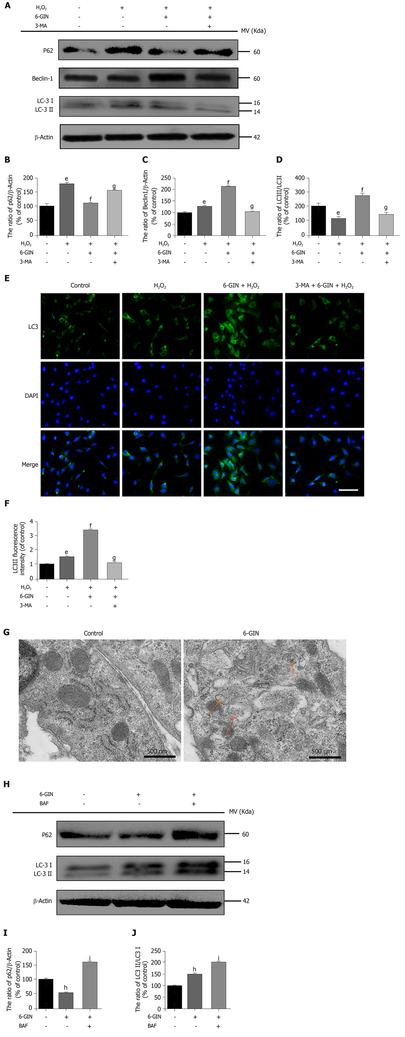
Figure 6 Role of autophagy in the protective effect of 6-gingerol.
Nucleus pulposus-derived mesenchymal stem cells (NPMSCs) were treated with cell culture medium (CON group), 80 μmol/L hydrogen peroxide alone (HYD group), hydrogen peroxide and 30 μmol/L 6-gingerol (6-GIN group), or hydrogen peroxide and 6-GIN combined with 10 mmol/L 3-methyladenine (3-MA group). A-D: Western blot results and quantitative analysis of the expression of the autophagy-related proteins P62, Beclin1, and LC3II/I; E-F: Immunofluorescence results and quantitative analysis of LC-3 expression (bar = 100 μm); G: Autophagosomes detected by transmission electron microscopy in NPMSCs. (Orange arrows: autophagosomes); H-J: Western blot results and quantitative analysis of the autophagy-related proteins P62 and LC3II/I in NPMSCs treated with 6-GIN (6-GIN group) or 6-GIN and 100 nM BAF (BAF group). All data are the mean ± SE of at least three independent experiments. hP < 0.05 vs blank control; iP < 0.05 vs 6-GIN only group. 6-GIN: 6-gingerol; H2O2: Hydrogen peroxide; 3-MA: 3-methyladenine.
- Citation: Nan LP, Wang F, Liu Y, Wu Z, Feng XM, Liu JJ, Zhang L. 6-gingerol protects nucleus pulposus-derived mesenchymal stem cells from oxidative injury by activating autophagy. World J Stem Cells 2020; 12(12): 1603-1622
- URL: https://www.wjgnet.com/1948-0210/full/v12/i12/1603.htm
- DOI: https://dx.doi.org/10.4252/wjsc.v12.i12.1603


