Copyright
©The Author(s) 2020.
World J Stem Cells. Dec 26, 2020; 12(12): 1576-1590
Published online Dec 26, 2020. doi: 10.4252/wjsc.v12.i12.1576
Published online Dec 26, 2020. doi: 10.4252/wjsc.v12.i12.1576
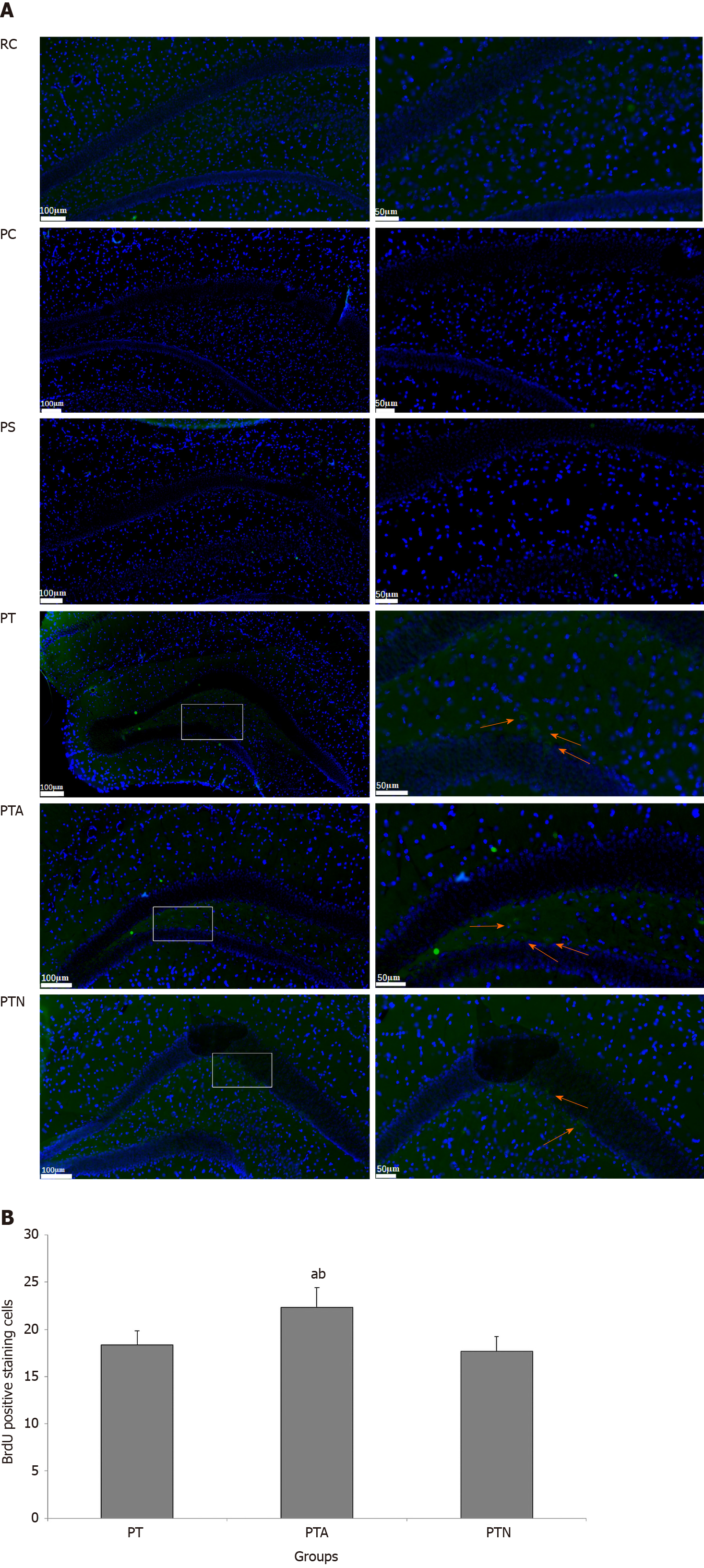
Figure 3 Neural stem cell proliferation by immunofluorescence staining.
Paraffin section of hippocampus tissue of mice was evaluated by indirect immunofluorescence and diaminobenzidine staining. After nuclei were counterstained with hematoxylin, positive BrdU-labeled cells were observed and counted. Arrows indicate positive cells marked by BrdU expression. A: Immunofluorescence staining; B: BrdU positive cell counting. aP < 0.05 when compared to senescence-accelerated mouse prone 8 (SAMP8) neural stem cells (NSCs) transplantation group, bP < 0.05 when compared to SAMP8 NSCs transplantation with non-acupoint group. RC: Senescence-accelerated mouse resistant 1 control group; PC: Senescence-accelerated mouse prone 8 control group; PS: Senescence-accelerated mouse prone 8 sham operation group; PT: Senescence-accelerated mouse prone 8 neural stem cells transplantation group; PTA: Senescence-accelerated mouse prone 8 neural stem cells transplantation with acupuncture group; PTN: Senescence-accelerated mouse prone 8 neural stem cells transplantation with non-acupoint group.
- Citation: Zhao L, Liu JW, Kan BH, Shi HY, Yang LP, Liu XY. Acupuncture accelerates neural regeneration and synaptophysin production after neural stem cells transplantation in mice. World J Stem Cells 2020; 12(12): 1576-1590
- URL: https://www.wjgnet.com/1948-0210/full/v12/i12/1576.htm
- DOI: https://dx.doi.org/10.4252/wjsc.v12.i12.1576


