Copyright
©The Author(s) 2020.
World J Stem Cells. Dec 26, 2020; 12(12): 1492-1510
Published online Dec 26, 2020. doi: 10.4252/wjsc.v12.i12.1492
Published online Dec 26, 2020. doi: 10.4252/wjsc.v12.i12.1492
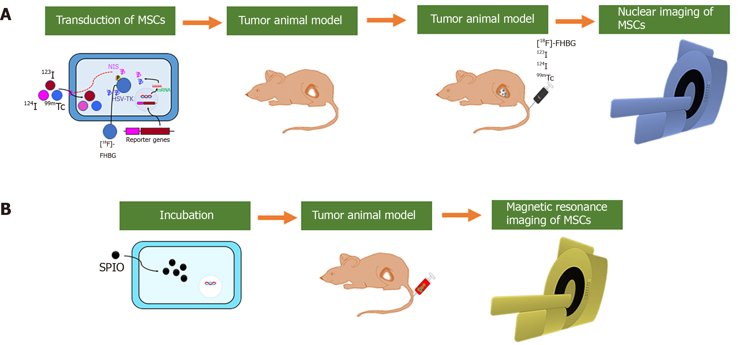
Figure 3 Schematic illustration of the labeling strategy for in vivo tracking of mesenchymal stem cells by nuclear and magnetic resonance imaging.
A: Gene transduction of sodium iodide symporter (NIS) or herpes simplex virus-thymidine kinase (HSV-TK) into mesenchymal stem cells (MSCs) can aid radiotracers (123I, 124I and 99mTc) in entering MSCs. MSCs-NIS are injected into tumor-bearing mice followed by the injection of radiotracers. In vivo nuclear imaging (positron-emission tomography, camera imaging, and single-photon emission computed tomography) can visualize migration of the MSCs; B: MSCs can be incubated with molecules including small superparamagnetic iron oxide (SPIO) or SPIO coated with gold-nanoparticles (SPIO@Au-NPs). SPIO-labeled MSCs are injected into tumor-bearing mice, and in vivo magnetic resonance imaging can visualize migration of the MSCs. MSCs: Mesenchymal stem cells; NIS: Sodium iodide symporter; HSV-TK: Herpes simplex virus-thymidine kinase; 99mTc: Technetium-99m; [18F]FHBG: 9-(4-[F]fluoro-3-hydroxymethylbutyl) guanine; SPIO: Superparamagnetic iron oxide.
- Citation: Rajendran RL, Jogalekar MP, Gangadaran P, Ahn BC. Noninvasive in vivo cell tracking using molecular imaging: A useful tool for developing mesenchymal stem cell-based cancer treatment. World J Stem Cells 2020; 12(12): 1492-1510
- URL: https://www.wjgnet.com/1948-0210/full/v12/i12/1492.htm
- DOI: https://dx.doi.org/10.4252/wjsc.v12.i12.1492


