Copyright
©The Author(s) 2020.
World J Stem Cells. Dec 26, 2020; 12(12): 1492-1510
Published online Dec 26, 2020. doi: 10.4252/wjsc.v12.i12.1492
Published online Dec 26, 2020. doi: 10.4252/wjsc.v12.i12.1492
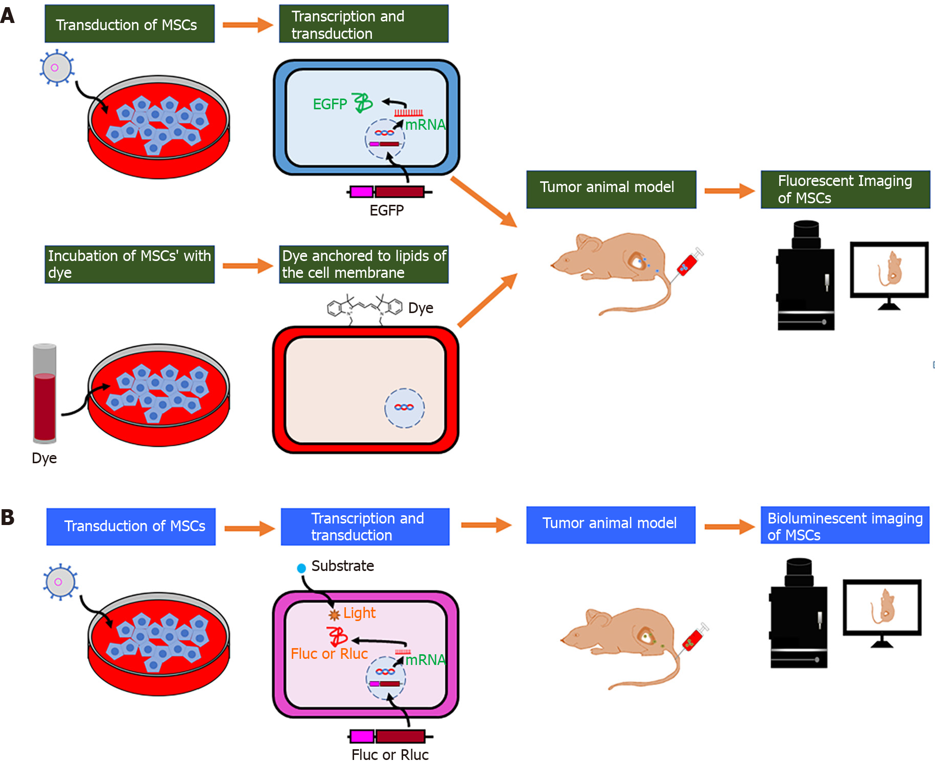
Figure 2 Schematic illustration of the labeling strategy for in vivo tracking of mesenchymal stem cells by optical imaging.
A: After fluorescent protein (enhanced green fluorescent protein) transduction into mesenchymal stem cells (MSCs) or binding of lipophilic labeling agents (e.g., fluorescent nanoparticles and VivoTrack 680) to the membrane of MSCs, cells are injected into the tumor-bearing mice, and their migration is visualized with the use of in vivo fluorescent imaging; B: After the bioluminescent protein (Firefly or Renilla luciferase) transduction into MSCs, cells are injected into the tumor-bearing mice. The light emitted due to the interaction between luciferase and its substrates (D-luciferin or coelenterazine) is captured by in vivo bioluminescent imaging. MSCs: Mesenchymal stem cells; EGFP: Enhanced green fluorescent protein; Fluc: Firefly luciferase; Rluc: Renilla luciferase.
- Citation: Rajendran RL, Jogalekar MP, Gangadaran P, Ahn BC. Noninvasive in vivo cell tracking using molecular imaging: A useful tool for developing mesenchymal stem cell-based cancer treatment. World J Stem Cells 2020; 12(12): 1492-1510
- URL: https://www.wjgnet.com/1948-0210/full/v12/i12/1492.htm
- DOI: https://dx.doi.org/10.4252/wjsc.v12.i12.1492


