Copyright
©The Author(s) 2020.
World J Stem Cells. Dec 26, 2020; 12(12): 1439-1454
Published online Dec 26, 2020. doi: 10.4252/wjsc.v12.i12.1439
Published online Dec 26, 2020. doi: 10.4252/wjsc.v12.i12.1439
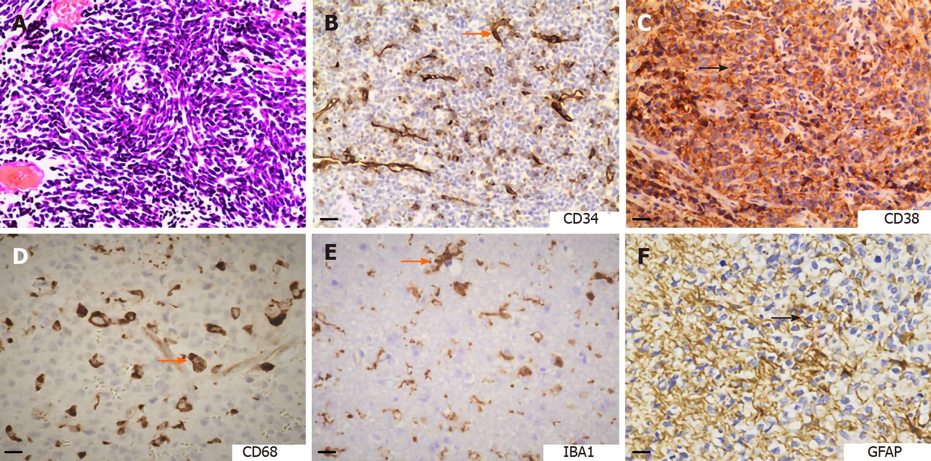
Figure 2 Hematoxylin and eosin staining and immunohistochemical staining of brain tumors.
A: Hematoxylin and eosin staining displaying the pathological vessels distributed in medulloblastoma (bar, 20 µm); B: Expression of CD34 (vascular endothelial cell marker) in chordoma (bar, 20 µm); C: Expression of CD38 (T cell marker) in anaplastic diffuse astrocytoma (bar, 20 µm); D: Expression of CD68 (macrophage marker) in medulloblastoma (bar, 10 µm); E: Expression of IBA1 (microglia marker) in glioblastoma (bar, 10 µm); F: Expression of glial fibrillary acidic protein (astrocyte marker) in medulloblastoma (bar, 10 µm). GFAP: Glial fibrillary acidic protein.
- Citation: Liu HL, Wang YN, Feng SY. Brain tumors: Cancer stem-like cells interact with tumor microenvironment. World J Stem Cells 2020; 12(12): 1439-1454
- URL: https://www.wjgnet.com/1948-0210/full/v12/i12/1439.htm
- DOI: https://dx.doi.org/10.4252/wjsc.v12.i12.1439


