Copyright
©The Author(s) 2020.
World J Stem Cells. Nov 26, 2020; 12(11): 1429-1438
Published online Nov 26, 2020. doi: 10.4252/wjsc.v12.i11.1429
Published online Nov 26, 2020. doi: 10.4252/wjsc.v12.i11.1429
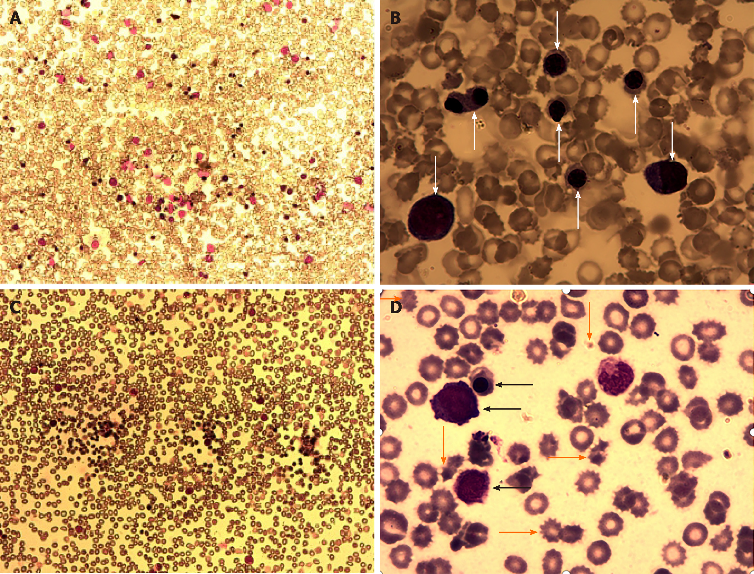
Figure 2 Morphological examination of bone marrow and periphery blood smears.
A: Morphological examination of bone marrow (BM) smears under low power lens (10 × 10) showed a normal cellularity, in the absence of fatty replacement; B: Morphological enumeration of BM nucleated cells under high power lens (10 × 100) showed an increased percentage of nucleated erythrocytes in multiple stages (marked by white arrows), with markedly reduced percentages of myeloid precursors and lymphocytes; C: Morphological examination of periphery blood (PB) smears under low power lens (10 × 10) showed an increase in nucleated cells, predominantly nucleated erythrocytes; D: Morphological enumeration of PB nucleated cells under high power lens (10 × 100) showed the presence of nucleated erythrocytes in multiple stages (marked by black arrows), with marked anisocytosis, acanthrocyte, and schistocyte in mature erythrocytes (marked by orange arrows). The morphological features of the BM and PB fulfilled the diagnosis of an erythroid proliferative disease and the toxic damage of erythrocytes.
- Citation: Zhao XC, Sun XY, Ju B, Meng FJ, Zhao HG. Acquired aplastic anemia: Is bystander insult to autologous hematopoiesis driven by immune surveillance against malignant cells? World J Stem Cells 2020; 12(11): 1429-1438
- URL: https://www.wjgnet.com/1948-0210/full/v12/i11/1429.htm
- DOI: https://dx.doi.org/10.4252/wjsc.v12.i11.1429


