Copyright
©The Author(s) 2020.
World J Stem Cells. Nov 26, 2020; 12(11): 1410-1428
Published online Nov 26, 2020. doi: 10.4252/wjsc.v12.i11.1410
Published online Nov 26, 2020. doi: 10.4252/wjsc.v12.i11.1410
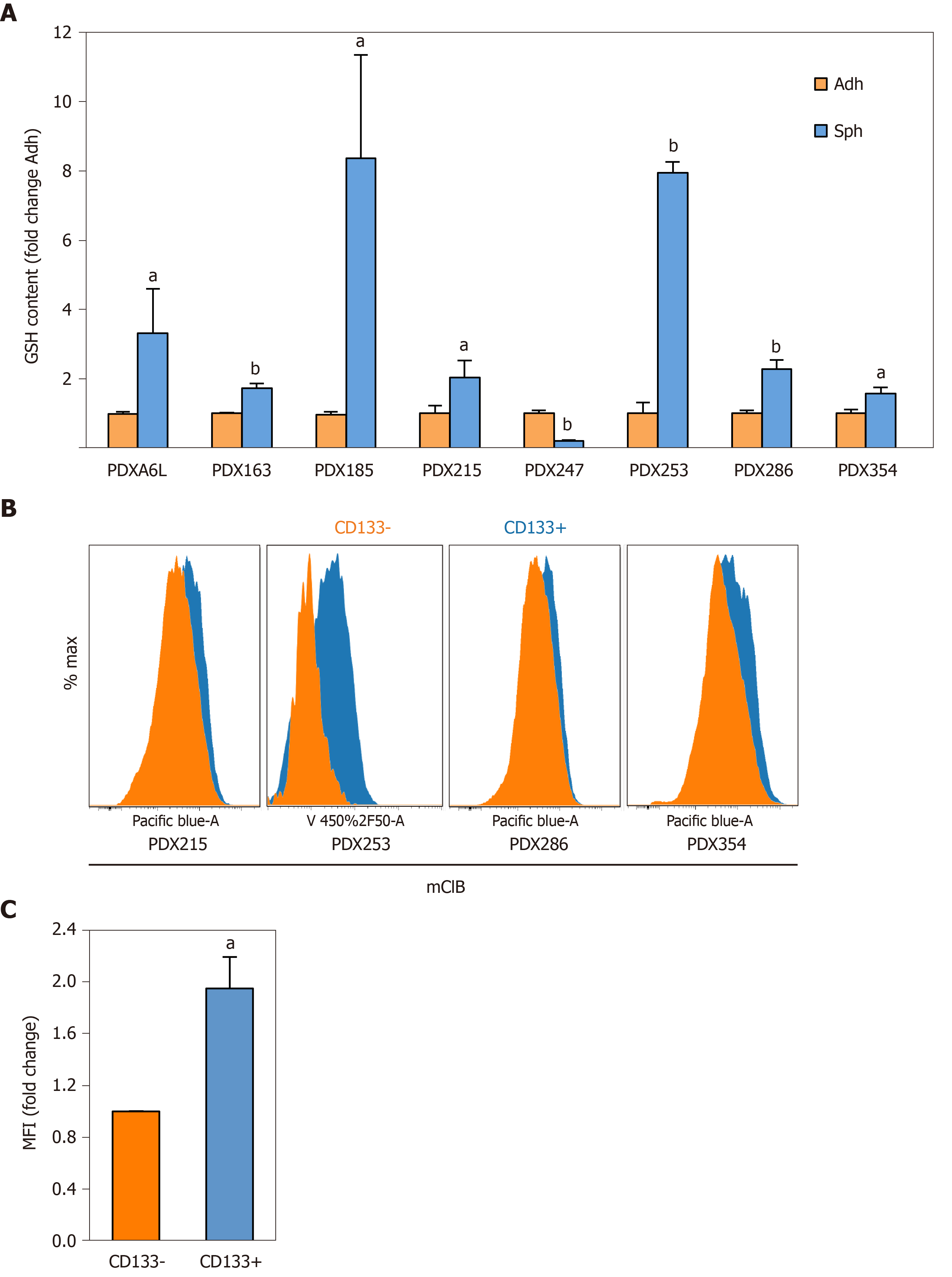
Figure 3 Reduced glutathione content is increased in cancer stem cell-enriching conditions.
Reduced glutathione (GSH) content was measured using the fluorescent thiol-reactive probe monochlorobimane (mClB). A: GSH content in cellular lysates was assessed by fluorimetry. Primary cells from different patient-derived xenograft (PDX) models as indicated in the figure were cultured in adherent or low-attachment cancer stem cell-enriching conditions for 7 d. Data were normalized for protein content; B: GSH content in CD133 positive and negative subpopulations as determined by flow cytometry. Representative flow cytometry histograms of the indicated PDX models are shown, with the following mean fluorescence intensities (MFI) for CD133– and CD133+ populations, respectively: PDX215 (2787 vs 4880), PDX286 (2748 vs 4364), PDX354 (4138 vs 6988); C: Pooled MFI data from PDX215, 286 and 354. Data in A and C are shown as mean ± SE fold change for sphere vs adherent cultures (A) or CD133+ vs CD133–. aP < 0.05; bP < 0.01. GSH: Glutathione content in its reduced form; PDX: Patient-derived xenograft; Sph: Sphere culture; Adh: adherent culture; MFI: Mean fluorescence intensities.
- Citation: Jagust P, Alcalá S, Sainz Jr B, Heeschen C, Sancho P. Glutathione metabolism is essential for self-renewal and chemoresistance of pancreatic cancer stem cells. World J Stem Cells 2020; 12(11): 1410-1428
- URL: https://www.wjgnet.com/1948-0210/full/v12/i11/1410.htm
- DOI: https://dx.doi.org/10.4252/wjsc.v12.i11.1410


