Copyright
©The Author(s) 2020.
World J Stem Cells. Nov 26, 2020; 12(11): 1327-1340
Published online Nov 26, 2020. doi: 10.4252/wjsc.v12.i11.1327
Published online Nov 26, 2020. doi: 10.4252/wjsc.v12.i11.1327
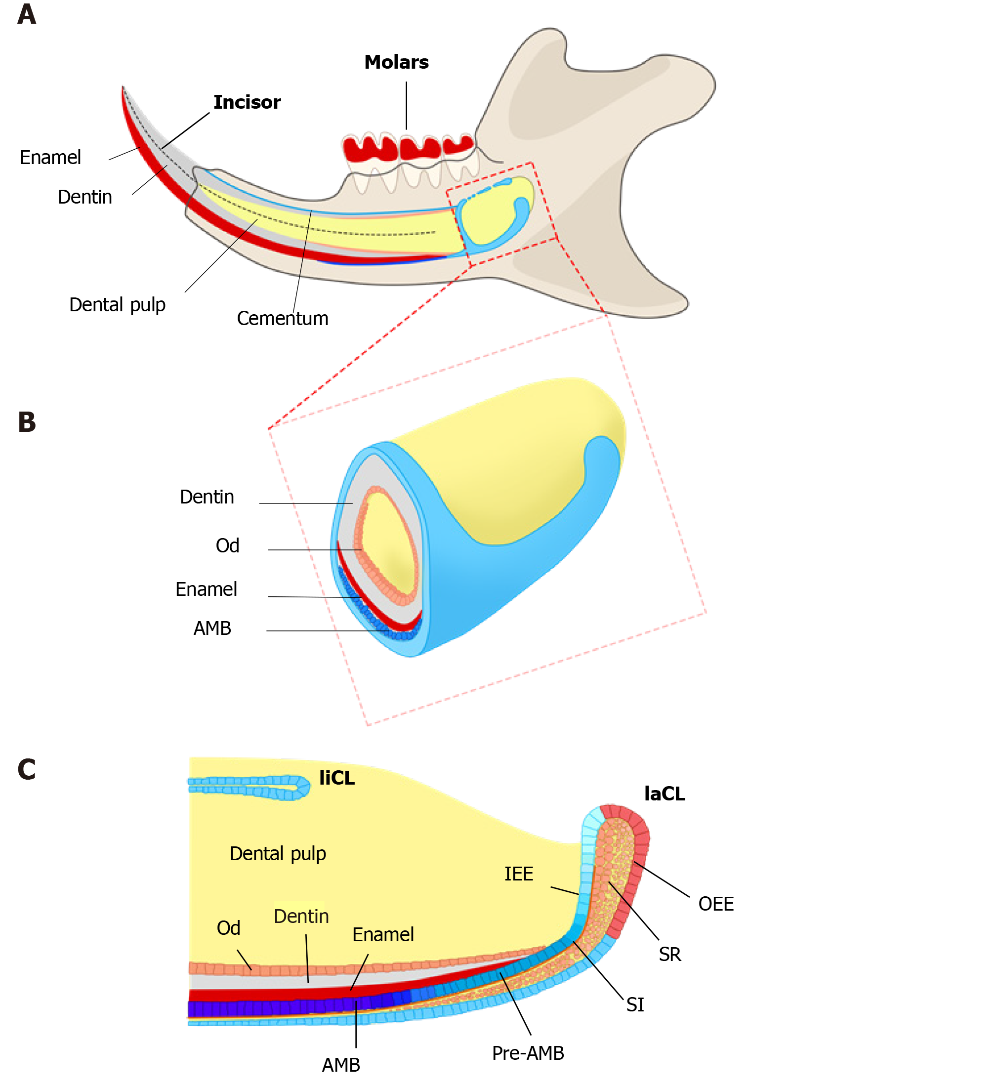
Figure 1 Schematics of adult mouse incisor and cervical loops.
A: Most of adult mouse incisor is embedded in the jawbone. Enamel (red) exists only at the labial side, which is produced by dental epithelial stem cells (DESCs) of the labial cervical loop (laCL). Dental pulp (yellow) is surrounded by dentin (gray), which is produced by dental mesenchymal stem cells. The lingual root analog is covered by cementum; B: Cervical loop is located in the proximal end of mouse incisor. The enamel and dentin are produced by the ameloblasts (AMB) and odontoblasts; C: Diagram of sagittal section of the proximal incisor. In growing adult mouse incisor, lingual cervical loops (liCL) and laCL persist throughout the adult stages. Slow-cycling DESCs are located in the outer enamel epithelium (OEE) and underlying stellate reticulum (SR). Transit-amplifying (TA) cells, also considered as actively cycling progenitors, undergo massive proliferation in the inner enamel epithelium. These cells give rise to pre-ameloblasts (Pre-AMB) and fully differentiated ameloblasts (AMB). Compared to the laCL, liCL is smaller and does not normally give rise to ameloblasts. SI: Stratum intermedium; AMB: Ameloblasts; OEE: Outer enamel epithelium; IEE: Inner enamel epithelium; SR: Stellate reticulum; Od: Odontoblasts; liCL: Lingual cervical loop; laCL: Labial cervical loop.
- Citation: Gan L, Liu Y, Cui DX, Pan Y, Wan M. New insight into dental epithelial stem cells: Identification, regulation, and function in tooth homeostasis and repair. World J Stem Cells 2020; 12(11): 1327-1340
- URL: https://www.wjgnet.com/1948-0210/full/v12/i11/1327.htm
- DOI: https://dx.doi.org/10.4252/wjsc.v12.i11.1327


