Copyright
©The Author(s) 2020.
World J Stem Cells. Oct 26, 2020; 12(10): 1152-1170
Published online Oct 26, 2020. doi: 10.4252/wjsc.v12.i10.1152
Published online Oct 26, 2020. doi: 10.4252/wjsc.v12.i10.1152
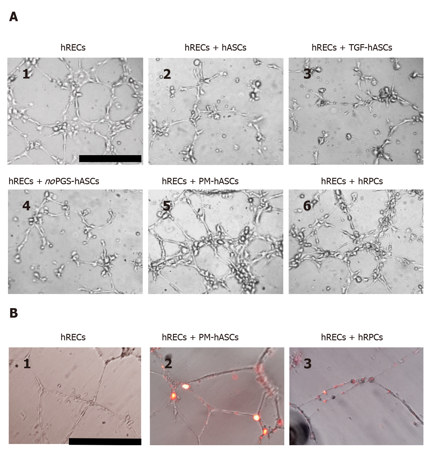
Figure 9 In vitro three-dimensional cell cultures in Matrigel.
A: After 6 h from seeding, human retinal endothelial cells (hRECs) cultured on Matrigel spontaneously form tubular microvessel-like structures (1). This predisposition appears enhanced when hRECs are co-cultured with human retinal pericyte cells (hRPCs) (6) or hASCs pre-cultured in complete pericyte medium (PM-hASCs) (5). No evident improvements are detectable if hRECs are co-cultured with control hASCs (2), hASCs pre-stimulated with transforming growth factor (3) or hASCs pre-cultured in pericyte medium lacking pericyte growth supplement (4). Scale bar: 100 μm. B: Typical tubular microvessel-like structures spontaneously formed by hRECs after 20 h from seeding (1). In 2 and 3, PM-hASCs and hRPCs, prelabeled with DiI (red fluorescence), were co-cultured with hRECs. In these cases, both cell types occupied the identical location in proximity to the tubular formations. Scale bar: 50 μm. hASCs: Human adipose derived mesenchymal stem cells pre-cultured in basal medium; hRECs: Human retinal endothelial cells; hRPCs: Human retinal pericyte cells; TGF-hASCs: hASCs pre-stimulated with transforming growth factor; noPGS-hASCs: hASCs pre-cultured in pericyte medium lacking pericyte growth supplement; PM-hASCs: hASCs pre-cultured in complete pericyte medium.
- Citation: Mannino G, Gennuso F, Giurdanella G, Conti F, Drago F, Salomone S, Lo Furno D, Bucolo C, Giuffrida R. Pericyte-like differentiation of human adipose-derived mesenchymal stem cells: An in vitro study. World J Stem Cells 2020; 12(10): 1152-1170
- URL: https://www.wjgnet.com/1948-0210/full/v12/i10/1152.htm
- DOI: https://dx.doi.org/10.4252/wjsc.v12.i10.1152


