Copyright
©The Author(s) 2020.
World J Stem Cells. Oct 26, 2020; 12(10): 1152-1170
Published online Oct 26, 2020. doi: 10.4252/wjsc.v12.i10.1152
Published online Oct 26, 2020. doi: 10.4252/wjsc.v12.i10.1152
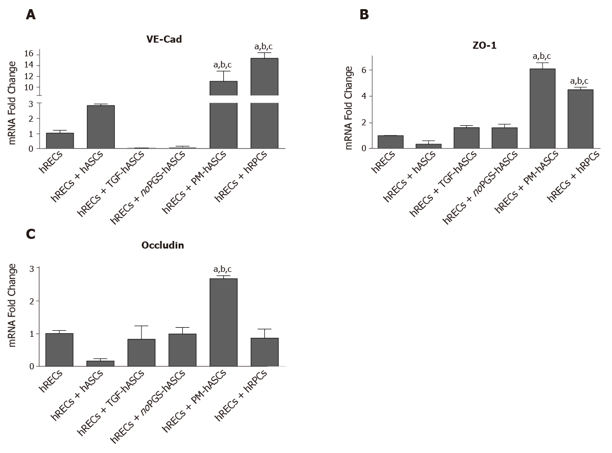
Figure 7 mRNA levels of junction proteins in human retinal endothelial cells after 4 days of co-culture with human retinal pericyte cells or different groups of human adipose-derived mesenchymal stem cells.
A: Histogram bars represent mRNA fold changes for vascular endothelial-Cadherin; B: Zonula occludens-1; and C: Occludin. Results are referred to the control levels of human retinal endothelial cells and normalized to the RNA expression of the housekeeping reference ribosomal gene 18S. All data represent mean ± SEM obtained from at least three independent experiments. Comparison between groups was evaluated by one-way ANOVA, followed by Tukey’s test. aIndicates significant difference (P < 0.05) vs human retinal endothelial cells; bIndicates significant difference (P < 0.05) vs human adipose-derived mesenchymal stem cells pre-stimulated with transforming growth factor; cIndicates significant difference (P < 0.05) vs human adipose-derived mesenchymal stem cells pre-cultured in pericyte medium lacking pericyte growth supplement.
- Citation: Mannino G, Gennuso F, Giurdanella G, Conti F, Drago F, Salomone S, Lo Furno D, Bucolo C, Giuffrida R. Pericyte-like differentiation of human adipose-derived mesenchymal stem cells: An in vitro study. World J Stem Cells 2020; 12(10): 1152-1170
- URL: https://www.wjgnet.com/1948-0210/full/v12/i10/1152.htm
- DOI: https://dx.doi.org/10.4252/wjsc.v12.i10.1152


