Copyright
©The Author(s) 2020.
World J Stem Cells. Oct 26, 2020; 12(10): 1152-1170
Published online Oct 26, 2020. doi: 10.4252/wjsc.v12.i10.1152
Published online Oct 26, 2020. doi: 10.4252/wjsc.v12.i10.1152
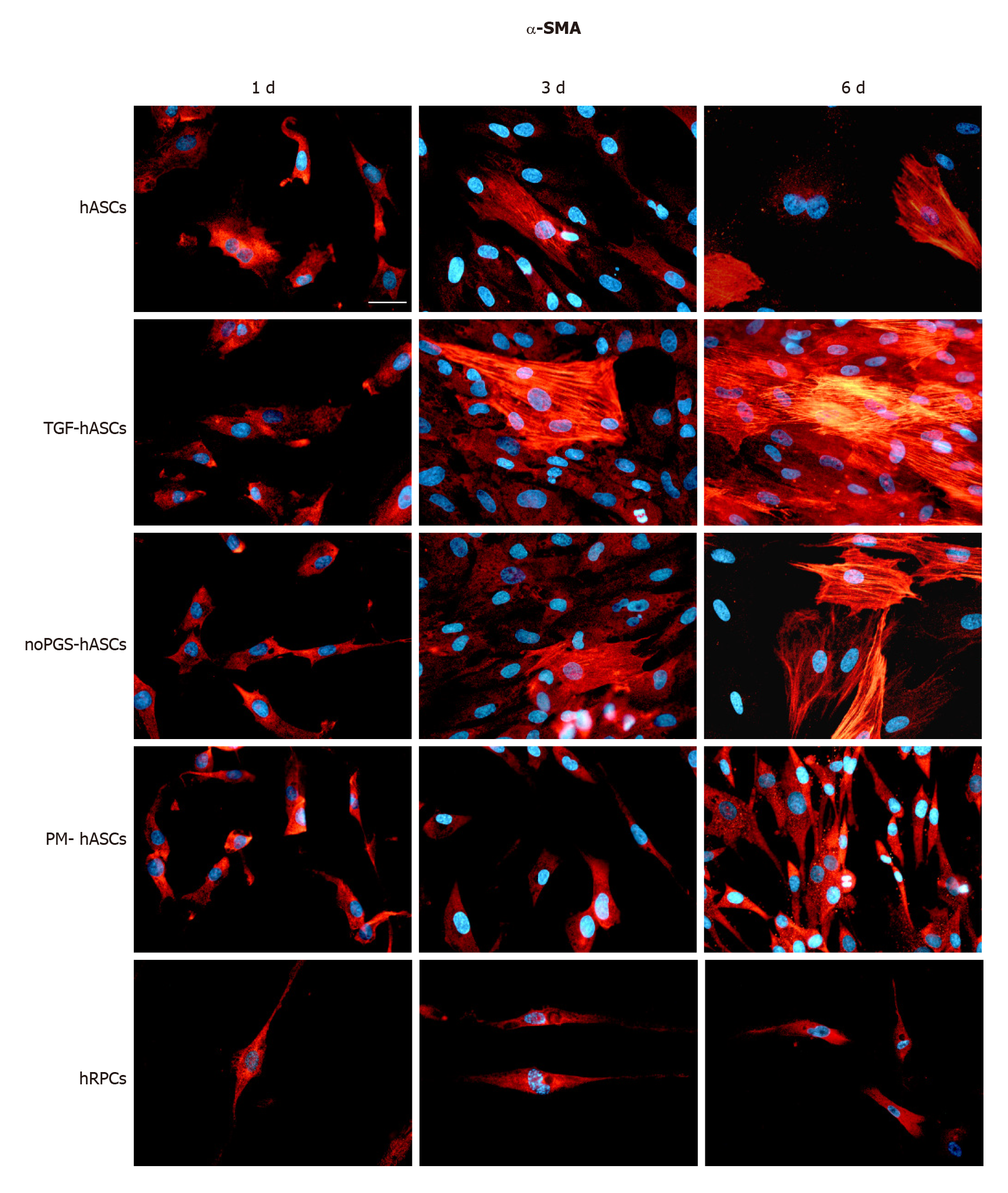
Figure 1 α-Smooth muscle actin immunoreactivity (red fluorescence) evaluated after one day (left column, 1 d), three days (middle column, 3 d) and six days (right column, 6 d) of cell growth.
First row: Human adipose-derived mesenchymal stem cells (hASCs) cultured in basal medium; Second row: hASCs in basal medium stimulated with transforming growth factor; Third row: hASCs cultured in pericyte medium without pericyte growth supplement; Fourth Row: hASCs cultured in complete pericyte medium; Fifth row: Human retinal pericyte cells cultured in complete pericyte medium (hRPCs). Photomicrographs show that only basal expression of α-SMA was detectable in hRPCs and all hASC groups at day 1 (left column). At day 3 and 6, a typical filamentous pattern of α-SMA was clearly detected in hASCs, hASCs in basal medium stimulated with transforming growth factor and hASCs cultured in pericyte medium without pericyte growth supplement, whereas the basal expression of α-SMA remained virtually unmodified in hASCs cultured in complete pericyte medium and hRPCs (last two rows). Blue fluorescence indicates DAPI staining of cell nuclei. Scale bar: 50 μm.
- Citation: Mannino G, Gennuso F, Giurdanella G, Conti F, Drago F, Salomone S, Lo Furno D, Bucolo C, Giuffrida R. Pericyte-like differentiation of human adipose-derived mesenchymal stem cells: An in vitro study. World J Stem Cells 2020; 12(10): 1152-1170
- URL: https://www.wjgnet.com/1948-0210/full/v12/i10/1152.htm
- DOI: https://dx.doi.org/10.4252/wjsc.v12.i10.1152


