Copyright
©The Author(s) 2020.
World J Stem Cells. Oct 26, 2020; 12(10): 1133-1151
Published online Oct 26, 2020. doi: 10.4252/wjsc.v12.i10.1133
Published online Oct 26, 2020. doi: 10.4252/wjsc.v12.i10.1133
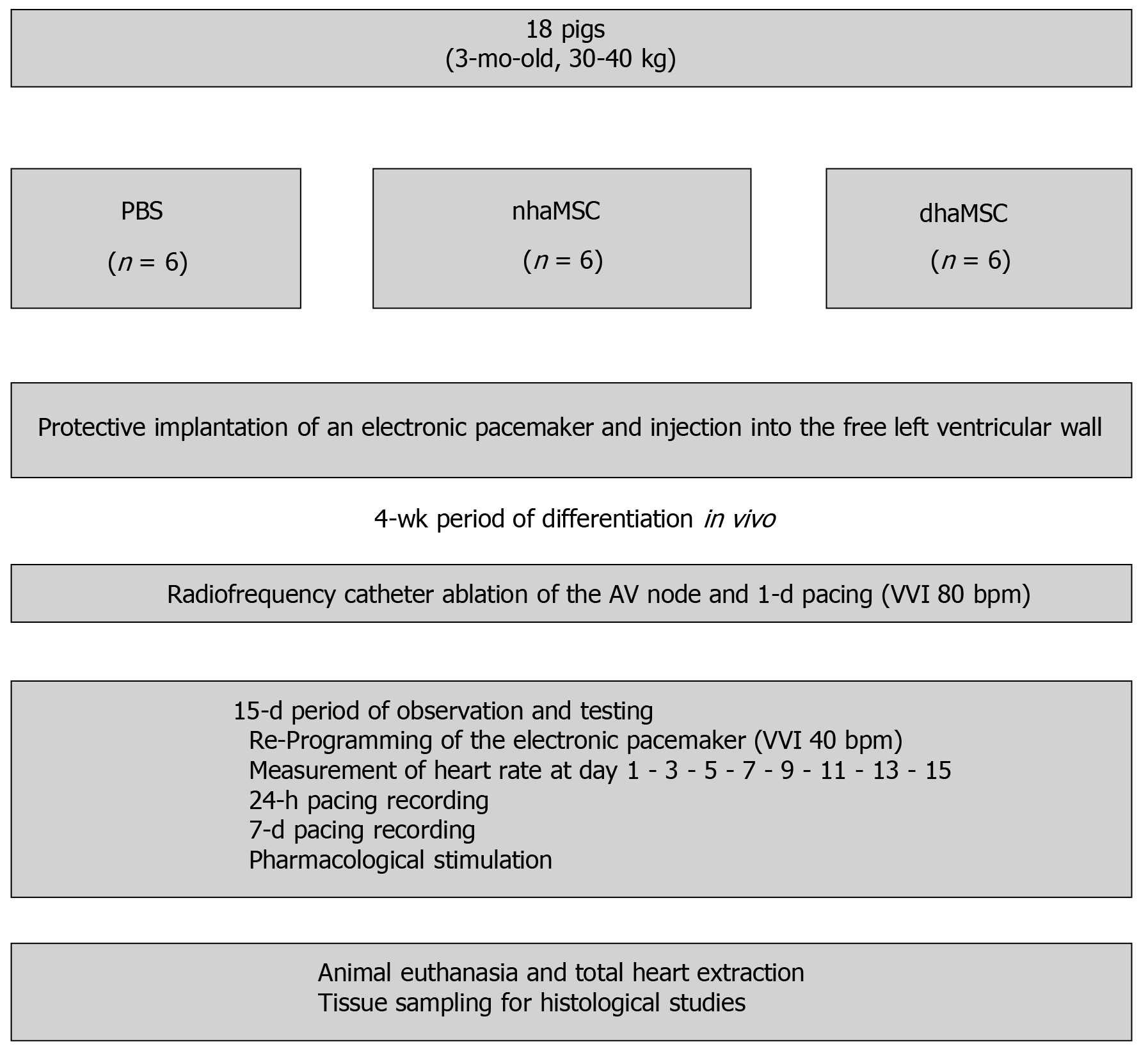
Figure 1 Schematic overview of in vivo experiments.
Eight healthy pigs (matched in sex, age and weight) were equally and randomly divided into 3 groups [phosphate-buffered saline (PBS), native human mesenchymal stem cells derived from adipose tissue, and differentiated human mesenchymal stem cells derived from adipose tissue]. Protective implantation of an electronic pacemaker was performed in each pig and cells or PBS were injected into the free left ventricular wall. After a 4-wk period, radiofrequency catheter ablation of the atrioventricular (AV) node was performed to establish third degree AV block in each animal. A 15-d period of observation and testing was followed by animal euthanasia and tissue sampling for histological studies. PBS: Phosphate-buffered saline; nhaMSC: Native human mesenchymal stem cells derived from adipose tissue; dhaMSC: Differentiated human mesenchymal stem cells derived from adipose tissue; AV: Atrioventricular.
- Citation: Darche FF, Rivinius R, Rahm AK, Köllensperger E, Leimer U, Germann G, Reiss M, Koenen M, Katus HA, Thomas D, Schweizer PA. In vivo cardiac pacemaker function of differentiated human mesenchymal stem cells from adipose tissue transplanted into porcine hearts. World J Stem Cells 2020; 12(10): 1133-1151
- URL: https://www.wjgnet.com/1948-0210/full/v12/i10/1133.htm
- DOI: https://dx.doi.org/10.4252/wjsc.v12.i10.1133


