Copyright
©The Author(s) 2019.
World J Stem Cells. Sep 26, 2019; 11(9): 705-721
Published online Sep 26, 2019. doi: 10.4252/wjsc.v11.i9.705
Published online Sep 26, 2019. doi: 10.4252/wjsc.v11.i9.705
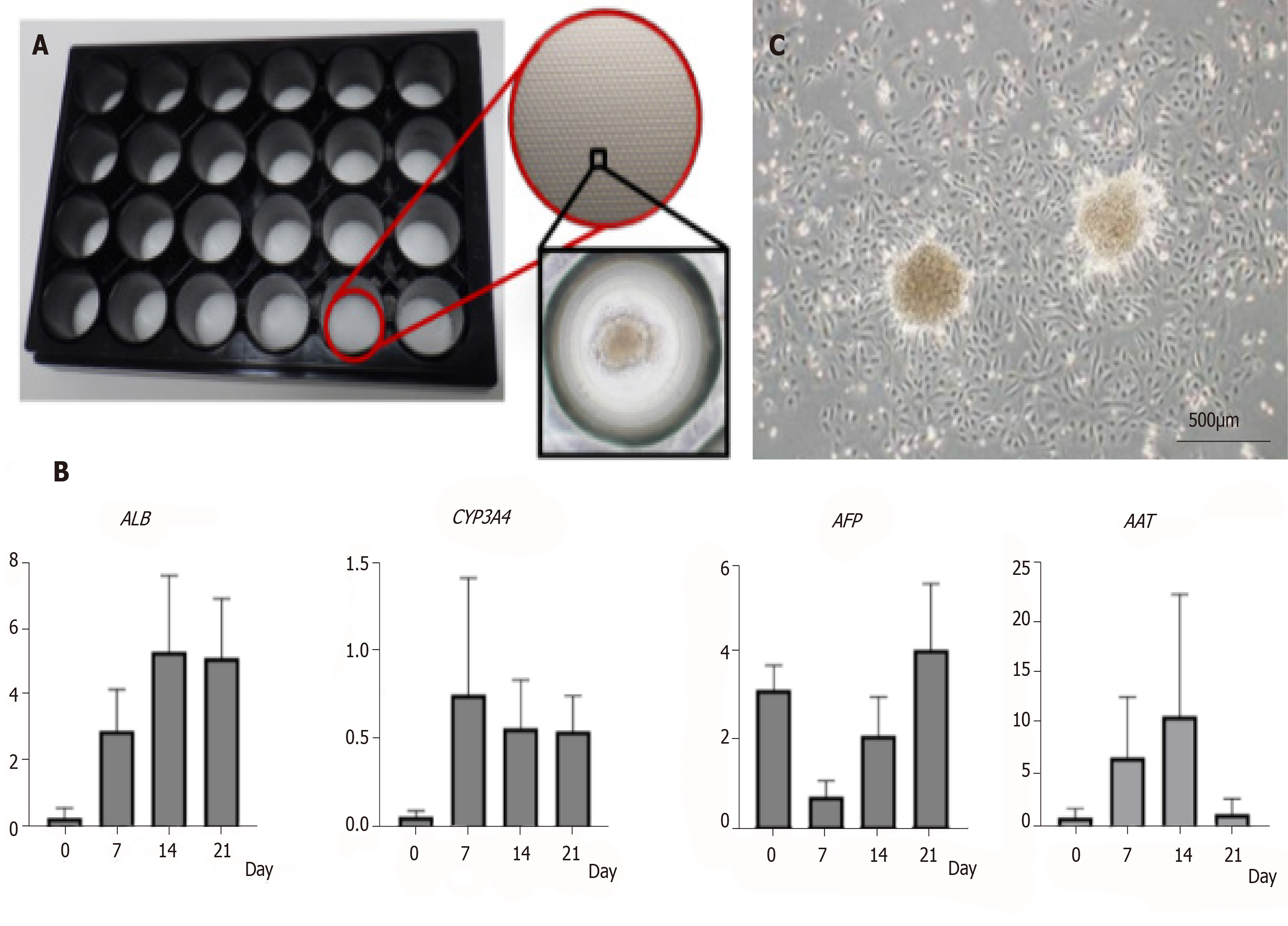
Figure 3 Characteristics of amniotic epithelial cell spheres formed on 3D-micropattern plate.
A: 3D-micropattern plate used in the present study. Round pits 500 µm in diameter are clustered on the surface. After culture, the amniotic epithelial cells (AECs) formed a sphere; B: Gene expression in the AEC sphere verified by qRT-PCR; C: After reseeding AEC sphere onto 2D culture dish, AEC proliferation was verified by phase-contrast microscopy. Bar, 500 µm.
- Citation: Furuya K, Zheng YW, Sako D, Iwasaki K, Zheng DX, Ge JY, Liu LP, Furuta T, Akimoto K, Yagi H, Hamada H, Isoda H, Oda T, Ohkohchi N. Enhanced hepatic differentiation in the subpopulation of human amniotic stem cells under 3D multicellular microenvironment. World J Stem Cells 2019; 11(9): 705-721
- URL: https://www.wjgnet.com/1948-0210/full/v11/i9/705.htm
- DOI: https://dx.doi.org/10.4252/wjsc.v11.i9.705


