Copyright
©The Author(s) 2019.
World J Stem Cells. Jul 26, 2019; 11(7): 383-397
Published online Jul 26, 2019. doi: 10.4252/wjsc.v11.i7.383
Published online Jul 26, 2019. doi: 10.4252/wjsc.v11.i7.383
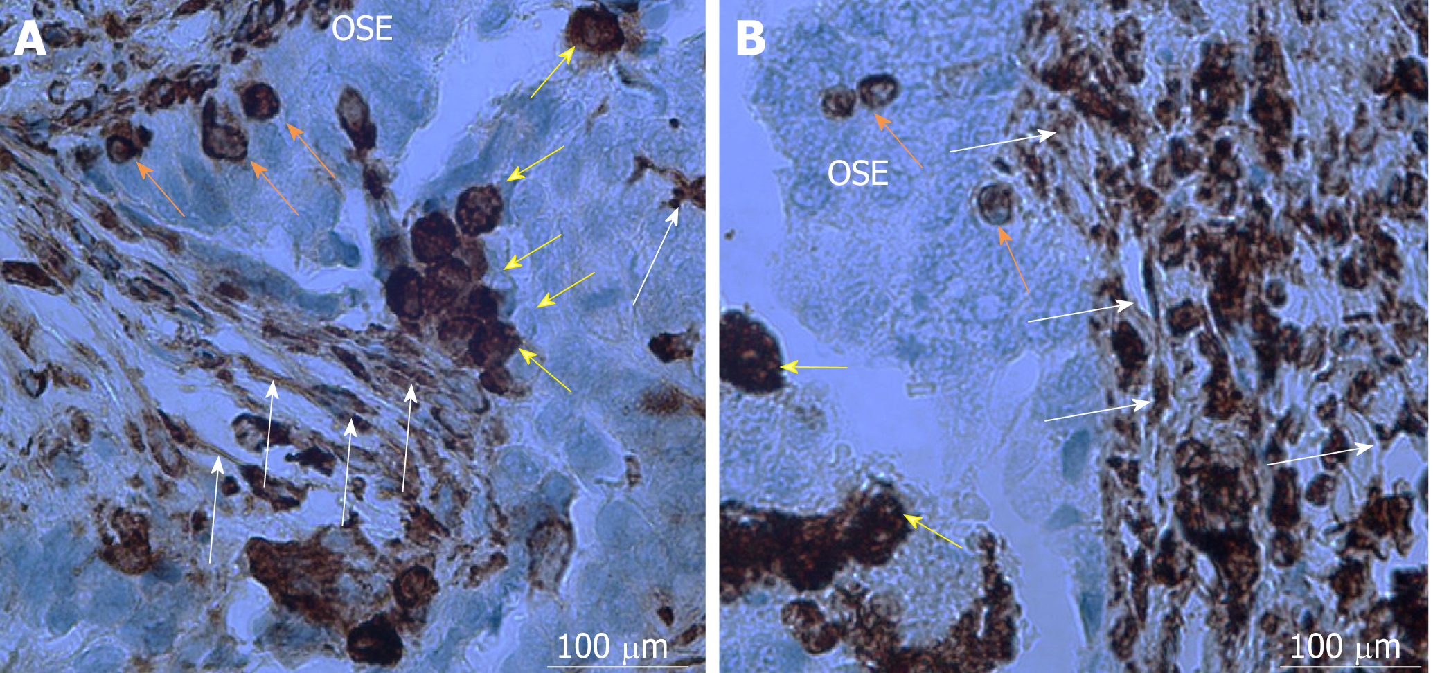
Figure 1 Cancer stem cells in ovarian cancer tissue sections after immunohistochemistry for vimentin.
A, B: Vimentin-positive small stem cells (≤ 5 μm) in ovarian surface epithelium (orange arrows, A and B) are developing into bigger round cells (10-15 μm) (yellow arrows, A and B) and mesenchymal-like stem cells (white arrows, A and B) by making elongations and protrusions. Legend: brown color-positivity for vimentin. Red bar: 100 μm.
- Citation: Kenda Suster N, Virant-Klun I. Presence and role of stem cells in ovarian cancer. World J Stem Cells 2019; 11(7): 383-397
- URL: https://www.wjgnet.com/1948-0210/full/v11/i7/383.htm
- DOI: https://dx.doi.org/10.4252/wjsc.v11.i7.383


