Copyright
©The Author(s) 2019.
World J Stem Cells. Feb 26, 2019; 11(2): 100-123
Published online Feb 26, 2019. doi: 10.4252/wjsc.v11.i2.100
Published online Feb 26, 2019. doi: 10.4252/wjsc.v11.i2.100
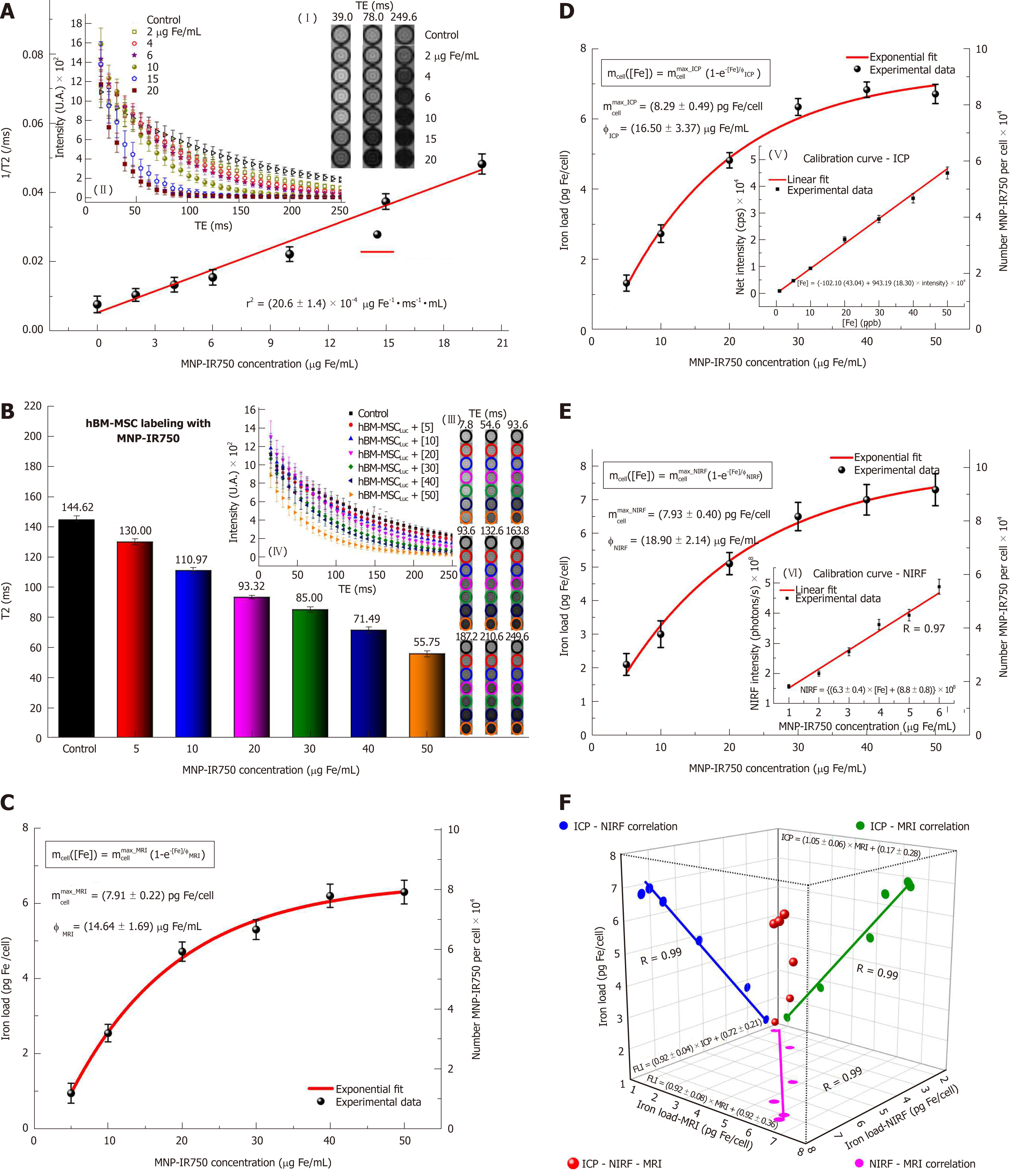
Figure 6 Quantification of multimodal nanoparticles-IR750 internalized by human bone marrow mesenchymal stem cellsLuc using magnetic resonance imaging, inductively coupled plasma-mass spectrometry and near-infrared fluorescence images in vitro.
For quantification via MRI, (A) the r2 values were determined from the intensity signal of MNP-IR750 at different concentrations as a function of TEs (I-II); B: the T2 values of hBM-MSCLuc labeled with MNP-IR750 at different concentrations were determined based on the adjustment of the MRI signal exponential decay curves obtained for the MRI images of ROIs (III-IV); C: the MNP-IR750 load and number internalized per cell as a function of different labeling concentrations were determined via MRI, and these values were adjusted based on an exponential curve, following equation 3 and the parameters determined for this equation; D: the MNP-IR750 load and number internalized per cell as a function of different labeling concentrations were determined via ICP-MS (V) from the calibration curve generated using known MNP-IR750 concentrations, and the values determined via ICP-MS quantification were adjusted based on an exponential curve, following equation 3 and the parameters determined for this equation; E: the MNP-IR750 load and number internalized per cell as a function of different labeling concentrations were determined via NIRF imaging (VI) from the calibration curve generated using known MNP-IR750 concentrations, and the values determined via NIRF quantification were adjusted with an exponential curve, following equation 3 and the parameters determined for this equation; and F: the correlation of the MNP-IR750 load internalized by hBM-MSCLuc was compared between ICP-MS and NIRF (blue dots), ICP-MS and MRI (green dots), NIRF and MRI (pink dots). hBM-MSC: Human bone marrow mesenchymal stem cells; MNP: Multimodal nanoparticles; ICP-MS: Inductively coupled plasma-mass spectrometry; MRI: Magnetic resonance imaging; NIRF: Near-infrared fluorescence; BLI: Bioluminescent images.
- Citation: da Silva HR, Mamani JB, Nucci MP, Nucci LP, Kondo AT, Fantacini DMC, de Souza LEB, Picanço-Castro V, Covas DT, Kutner JM, de Oliveira FA, Hamerschlak N, Gamarra LF. Triple-modal imaging of stem-cells labeled with multimodal nanoparticles, applied in a stroke model. World J Stem Cells 2019; 11(2): 100-123
- URL: https://www.wjgnet.com/1948-0210/full/v11/i2/100.htm
- DOI: https://dx.doi.org/10.4252/wjsc.v11.i2.100


