Copyright
©The Author(s) 2019.
World J Stem Cells. Feb 26, 2019; 11(2): 100-123
Published online Feb 26, 2019. doi: 10.4252/wjsc.v11.i2.100
Published online Feb 26, 2019. doi: 10.4252/wjsc.v11.i2.100
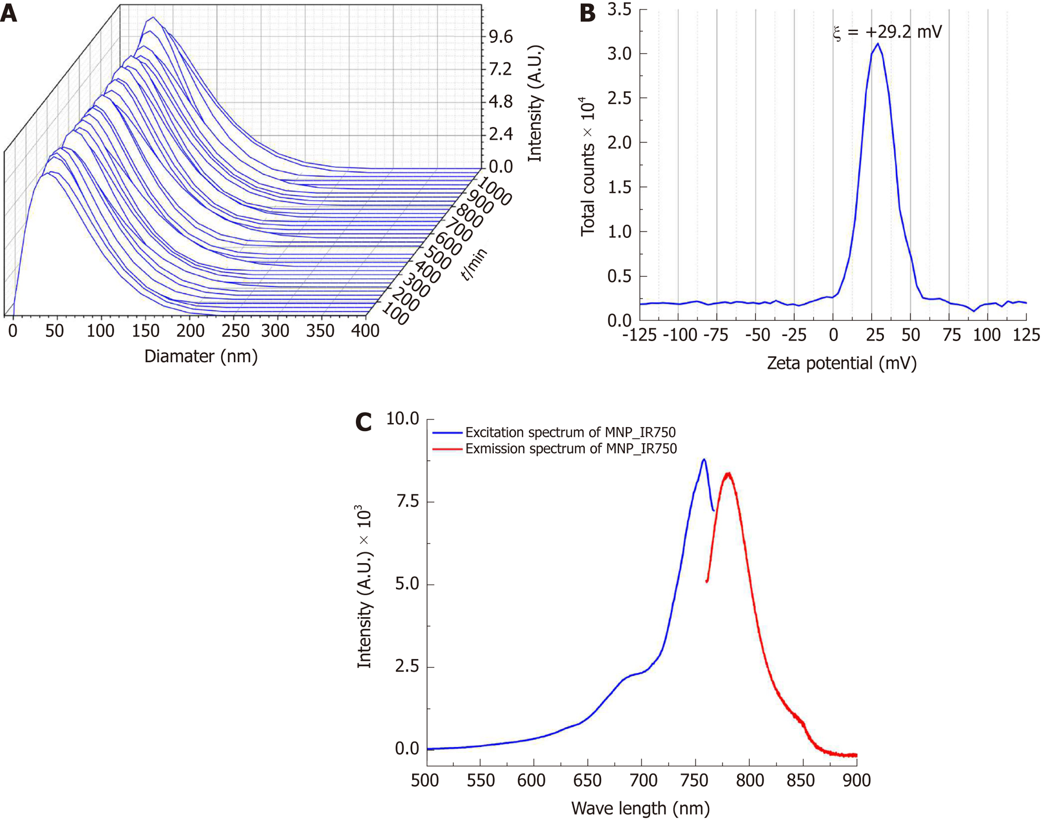
Figure 3 Characterization of the hydrodynamic diameter, zeta potential and optical properties of multimodal nanoparticles-IR750.
A: Curves of the hydrodynamic size distribution of MNP-IR750 over 18 h; B: Spectrum of the surface charge of MNP-IR750 with a zeta potential at pH 7.4 of ξ = +29.2±1.9 mV; C: MNP-IR750 excitation/emission spectrum (blue and red curves, respectively), showing fluorescence intensity peaks of 757.9 and 779.4 nm, respectively. MNP: Multimodal nanoparticles.
- Citation: da Silva HR, Mamani JB, Nucci MP, Nucci LP, Kondo AT, Fantacini DMC, de Souza LEB, Picanço-Castro V, Covas DT, Kutner JM, de Oliveira FA, Hamerschlak N, Gamarra LF. Triple-modal imaging of stem-cells labeled with multimodal nanoparticles, applied in a stroke model. World J Stem Cells 2019; 11(2): 100-123
- URL: https://www.wjgnet.com/1948-0210/full/v11/i2/100.htm
- DOI: https://dx.doi.org/10.4252/wjsc.v11.i2.100


