Copyright
©The Author(s) 2019.
World J Stem Cells. Nov 26, 2019; 11(11): 990-1004
Published online Nov 26, 2019. doi: 10.4252/wjsc.v11.i11.990
Published online Nov 26, 2019. doi: 10.4252/wjsc.v11.i11.990
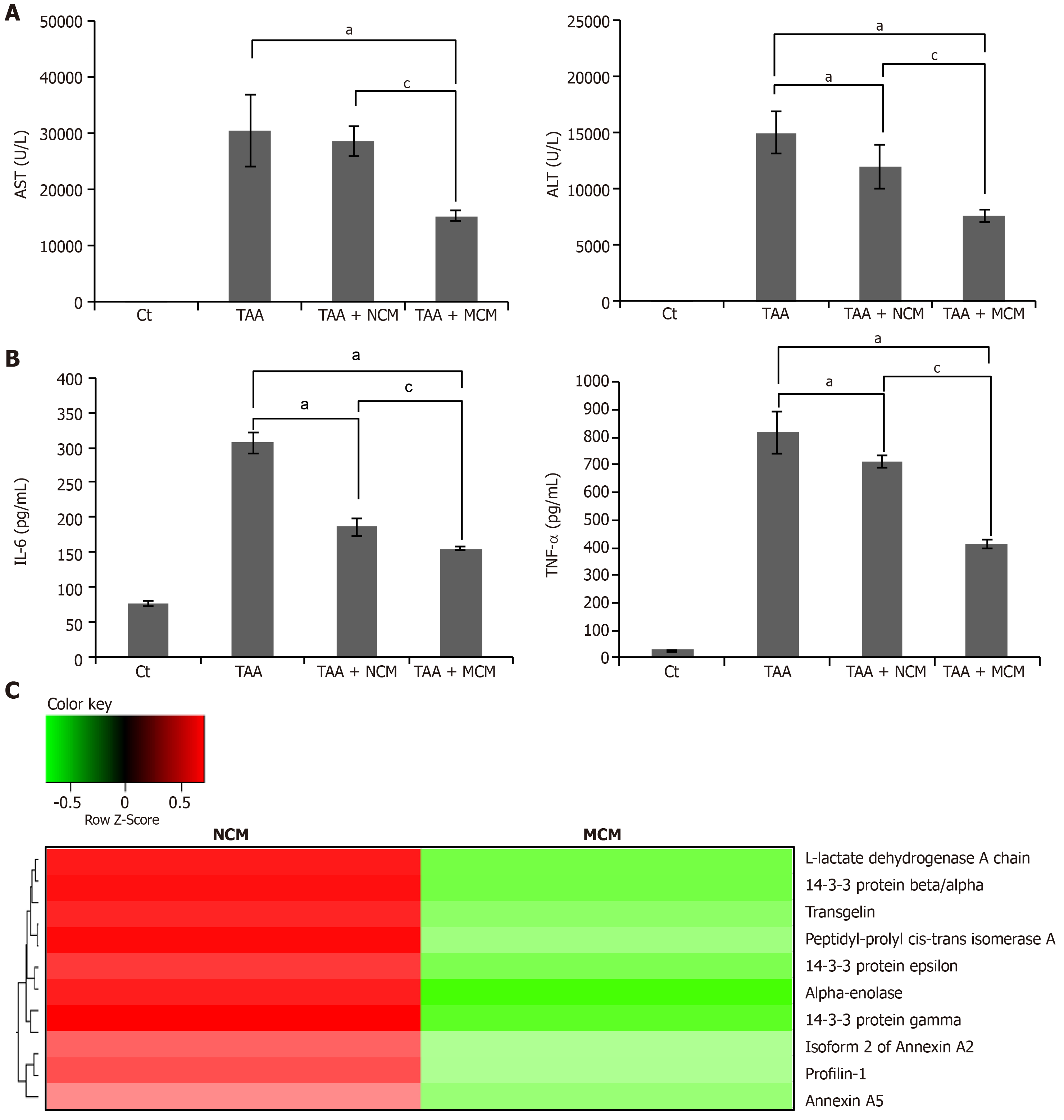
Figure 6 Determination of systemic effects of MCM and analysis of secretome components.
A: Results of ELISA showing serum levels of inflammatory markers (IL-6 and TNF-α) in each group. MCM administration had the greatest effect on lowering the serum levels of IL-6 and TNF-α in thioacetamide (TAA)-treated mice; B: Serology tests of AST and ALT in the mouse model of liver fibrosis. MCM infusion had the greatest effect on decreasing the serum levels of AST and ALT; C: Heat map generated from label-free LC-MS for quantitative proteomics reflecting protein expression values of NCM and MCM. Samples are arranged in columns, proteins in rows. Red shading indicates increased expression in samples compared to control; green shading indicates reduced expression; and black shading indicates median expression. Each sample for LC-MS was pooled from three samples of the secretome. The components and concentrations of various essential proteins varied widely between NCM and MCM, validating the effects of miR-125 transfection. Specifically, MCM exhibited a significantly lower concentration of essential intermediates of TGF-β/Smad signaling, such as transgelin, PIN1, and Profilin-1, than NCM. Values are presented as mean ± standard deviation of three independent experiments. aP < 0.05 vs Ct (TAA). cP < 0.05 between TAA + NCM and TAA + MCM. ALT: Alanine transaminase; AST: Aspartate transaminase; TAA: Thioacetamide; TNF- α: Tumor necrosis factor-α.
- Citation: Kim KH, Lee JI, Kim OH, Hong HE, Kwak BJ, Choi HJ, Ahn J, Lee TY, Lee SC, Kim SJ. Ameliorating liver fibrosis in an animal model using the secretome released from miR-122-transfected adipose-derived stem cells. World J Stem Cells 2019; 11(11): 990-1004
- URL: https://www.wjgnet.com/1948-0210/full/v11/i11/990.htm
- DOI: https://dx.doi.org/10.4252/wjsc.v11.i11.990


