Copyright
©The Author(s) 2019.
World J Stem Cells. Nov 26, 2019; 11(11): 990-1004
Published online Nov 26, 2019. doi: 10.4252/wjsc.v11.i11.990
Published online Nov 26, 2019. doi: 10.4252/wjsc.v11.i11.990
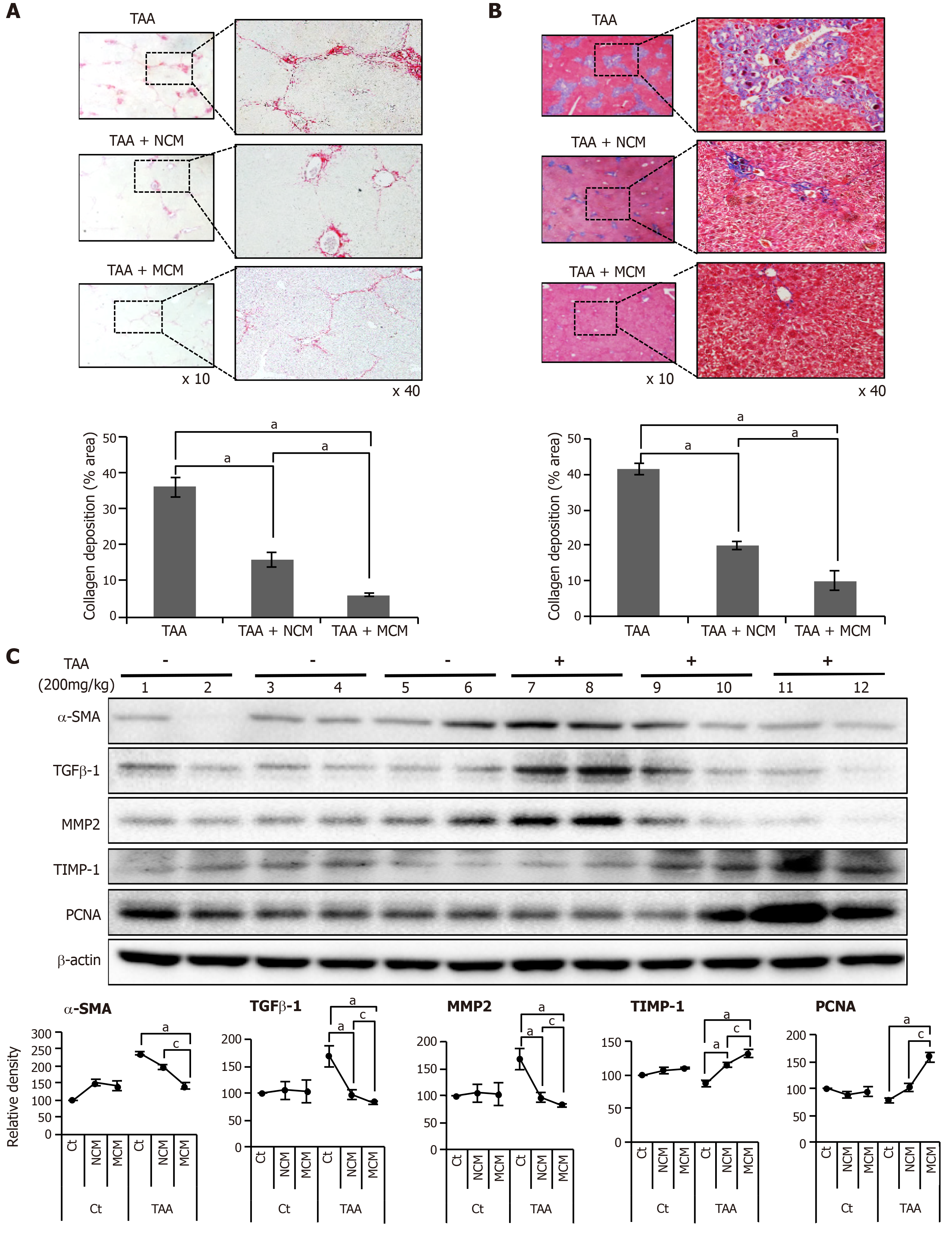
Figure 3 Determination of antifibrotic effects of MCM in the in vivo model of liver fibrosis.
Control mice and thioacetamide (TAA)-treated mice (mouse model of liver fibrosis) were intravenously (using tail vein) infused with normal saline, CM, and MCM. A, B: Sirius red A and Masson’s trichrome B stains showing that MCM infusion significantly decreased the collagen content of the liver in the mouse model of liver fibrosis. Magnification × 400. Percentages of fibrotic areas were measured using NIH image J and expressed as relative values to those in normal livers; C: Western blot analysis of liver specimens. MCM infusion significantly increased the expression of PCNA (a proliferation marker), and significantly decreased the expression of α-SMA, TGF-β1, and MMP1 (fibrotic markers) and increased an antifibrotic marker (TIMP-1) in the mouse model of liver fibrosis. The relative densities of individual markers had been quantified using Image Lab 3.0 (Bio-Rad) software and then were normalized to that of β-actin in each group. Values are presented as mean ± standard deviation of three independent experiments. aP < 0.05 vs Ct (TAA). cP < 0.05 between TAA + NCM and TAA + MCM. α-SMA: Alpha-smooth muscle actin; Ct: Control; CM: The secretome obtained from ASCs after 48-h-incubation; MCM: The secretome released from miR-122-transfected ASCs; MMP-1: Metalloproteinases-1; PCNA: Proliferating cell nuclear antigen; TAA: Thioacetamide; TGF-β: Transforming growth factor-β; TIMP-1: Tissue inhibitor of metalloproteinases-1.
- Citation: Kim KH, Lee JI, Kim OH, Hong HE, Kwak BJ, Choi HJ, Ahn J, Lee TY, Lee SC, Kim SJ. Ameliorating liver fibrosis in an animal model using the secretome released from miR-122-transfected adipose-derived stem cells. World J Stem Cells 2019; 11(11): 990-1004
- URL: https://www.wjgnet.com/1948-0210/full/v11/i11/990.htm
- DOI: https://dx.doi.org/10.4252/wjsc.v11.i11.990


