Copyright
©The Author(s) 2019.
World J Stem Cells. Jan 26, 2019; 11(1): 33-43
Published online Jan 26, 2019. doi: 10.4252/wjsc.v11.i1.33
Published online Jan 26, 2019. doi: 10.4252/wjsc.v11.i1.33
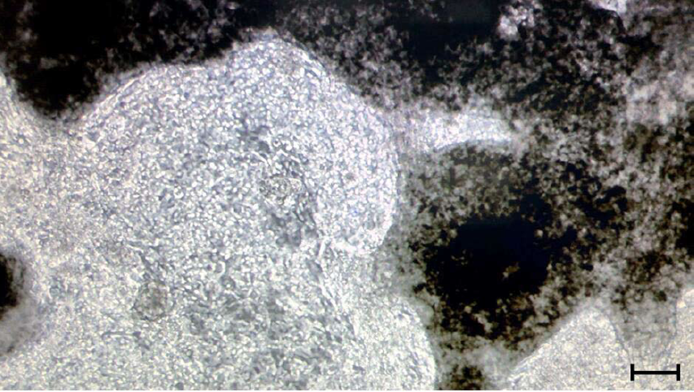
Figure 3 Induced pluripotent stem cell-derived cardiomyocytes in a 3-dimensional structure.
Phase contrast image of a 3D formation of induced pluripotent stem cell-derived cardiomyocytes. Control hiPSCs were differentiated into hiPSC-CMs using STEMCELL Technologies cardiomyocyte differentiation kit. Image was taken at day 14 of differentiation. Scale bar indicates 40 μm.
- Citation: Machiraju P, Greenway SC. Current methods for the maturation of induced pluripotent stem cell-derived cardiomyocytes. World J Stem Cells 2019; 11(1): 33-43
- URL: https://www.wjgnet.com/1948-0210/full/v11/i1/33.htm
- DOI: https://dx.doi.org/10.4252/wjsc.v11.i1.33


