Copyright
©The Author(s) 2018.
World J Stem Cells. Dec 26, 2018; 10(12): 212-227
Published online Dec 26, 2018. doi: 10.4252/wjsc.v10.i12.212
Published online Dec 26, 2018. doi: 10.4252/wjsc.v10.i12.212
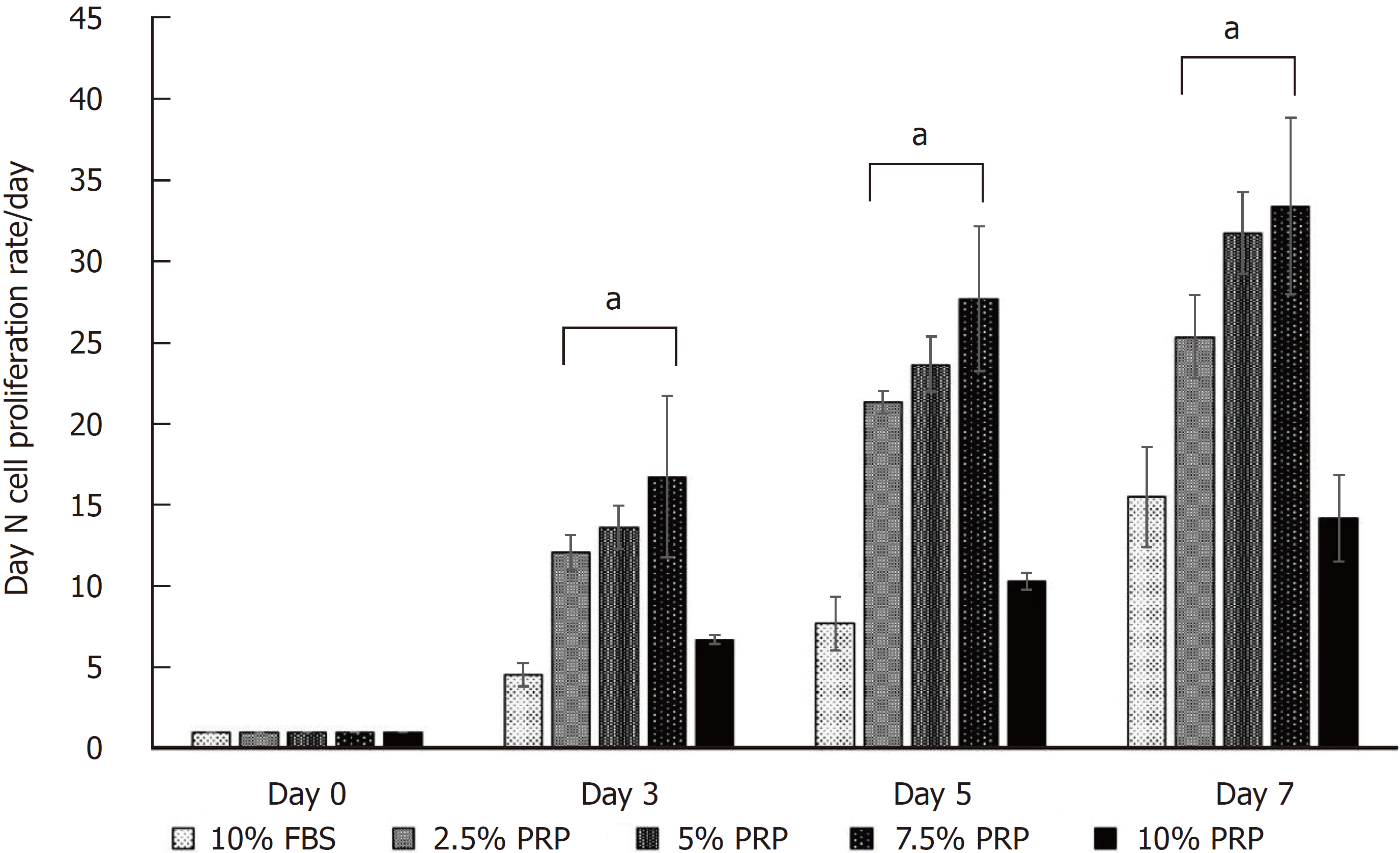
Figure 2 Cell proliferation assay.
At the endpoint (day 7), the proliferation rate of ADSCs was significantly higher in the 2.5%, 5%, and 7.5% PRP groups (25.348 ± 2.572, 31.778 ± 2.523, 33.400 ± 5.428 for 2.5% PRP, 5% PRP, 7.5% PRP, respectively) than in the 10% FBS (control group) and 10% PRP groups (15.483 ± 3.071 and 14.168 ± 2.650 for 10% FBS and 10% PRP; P < 0.01). The results suggested that 2.5%, 5%, and 7.5% PRP showed a higher ability to increase ADSC proliferation compared with FBS. Data are expressed as mean ± standard deviation. aP < 0.01 vs 10% FBS group. ADSC: Adipose-derived stem cell; PRP: Platelet-rich plasma; FBS: Fetal bovine serum.
- Citation: Chen CF, Liao HT. Platelet-rich plasma enhances adipose-derived stem cell-mediated angiogenesis in a mouse ischemic hindlimb model. World J Stem Cells 2018; 10(12): 212-227
- URL: https://www.wjgnet.com/1948-0210/full/v10/i12/212.htm
- DOI: https://dx.doi.org/10.4252/wjsc.v10.i12.212


