Copyright
©The Author(s) 2018.
World J Stem Cells. Nov 26, 2018; 10(11): 146-159
Published online Nov 26, 2018. doi: 10.4252/wjsc.v10.i11.146
Published online Nov 26, 2018. doi: 10.4252/wjsc.v10.i11.146
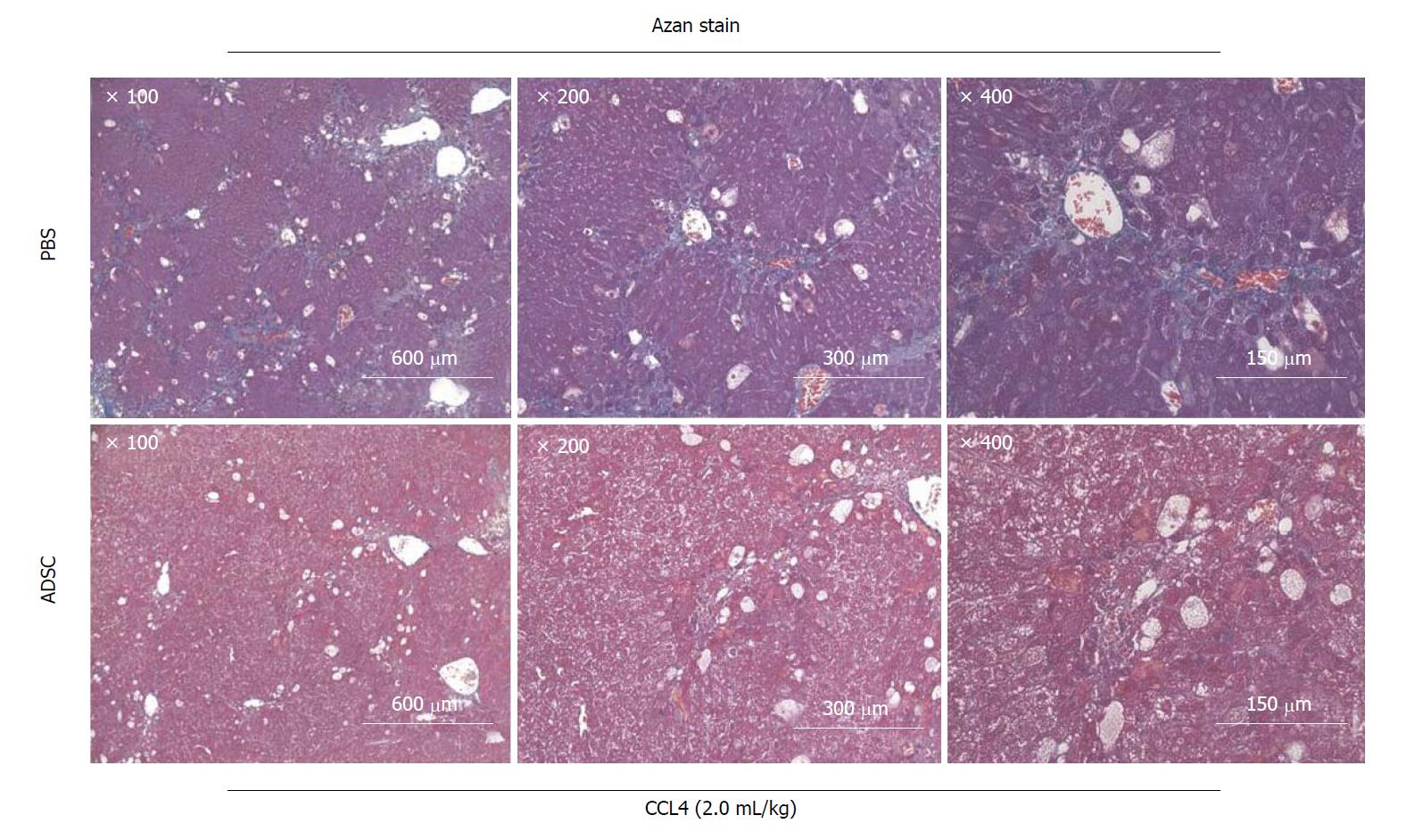
Figure 4 Adipose-derived mesenchymal stem cells improved symptoms of tissue fibrosis in cirrhosis caused by the administration of CCL4.
Micrographic image of Azan staining of liver specimens. Azan staining is a fibrous connective tissue staining method that differentiates collagen fibers and muscle fibers. The fibrous connective tissue in the tissue section was stained blue. Microscopic images of the liver specimens 20 h after the administration of PBS (upper panels) and adipose-derived mesenchymal stem cells (ADSC) (lower panels) via the mouse tail vein. In the ADSC administration group, fibrosis and pseudolobule formation were ameliorated. Microscopic images (×100 - 400) of the same tissue section.
- Citation: Nahar S, Nakashima Y, Miyagi-Shiohira C, Kinjo T, Toyoda Z, Kobayashi N, Saitoh I, Watanabe M, Noguchi H, Fujita J. Cytokines in adipose-derived mesenchymal stem cells promote the healing of liver disease. World J Stem Cells 2018; 10(11): 146-159
- URL: https://www.wjgnet.com/1948-0210/full/v10/i11/146.htm
- DOI: https://dx.doi.org/10.4252/wjsc.v10.i11.146


