Copyright
©The Author(s) 2018.
World J Stem Cells. Jan 26, 2018; 10(1): 1-14
Published online Jan 26, 2018. doi: 10.4252/wjsc.v10.i1.1
Published online Jan 26, 2018. doi: 10.4252/wjsc.v10.i1.1
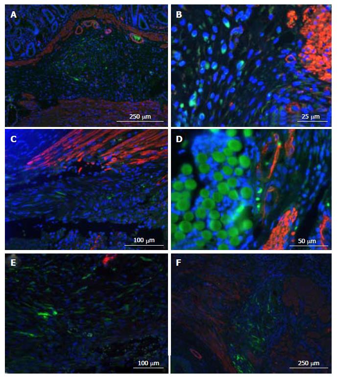
Figure 7 Immunolocalization of ASCs at surgical site.
Cell nuclei were stained with DAPI (blue), ASCs with eGFP (green), and muscle with α-actin (red). On injected animals, ASCs tended to form “conglomerates” in the submucosa and were able to survive at least 7 d (A: G1 group rat sacrificed at 7 d). ASCs appeared in injury foci, even though they were deposited at some distance (e.g., in “conglomerates”): In muscle section margins (B: G2 sacrificed at 7 d and C: G1 sacrificed at 1 d). In the biosuture group, ACSs also surround the suture (D: G2 sacrificed at 4 d). ASCs also appeared in the tissue that filled the space between muscle section margins (E: G3 group rat sacrificed at 4 d and F: G1 group rat sacrificed at 7 d). ASC: Adipose-derived stem cell; eGFP: Enhanced green fluorescent protein.
- Citation: Trébol J, Georgiev-Hristov T, Vega-Clemente L, García-Gómez I, Carabias-Orgaz A, García-Arranz M, García-Olmo D. Rat model of anal sphincter injury and two approaches for stem cell administration. World J Stem Cells 2018; 10(1): 1-14
- URL: https://www.wjgnet.com/1948-0210/full/v10/i1/1.htm
- DOI: https://dx.doi.org/10.4252/wjsc.v10.i1.1


