Copyright
©The Author(s) 2018.
World J Stem Cells. Jan 26, 2018; 10(1): 1-14
Published online Jan 26, 2018. doi: 10.4252/wjsc.v10.i1.1
Published online Jan 26, 2018. doi: 10.4252/wjsc.v10.i1.1
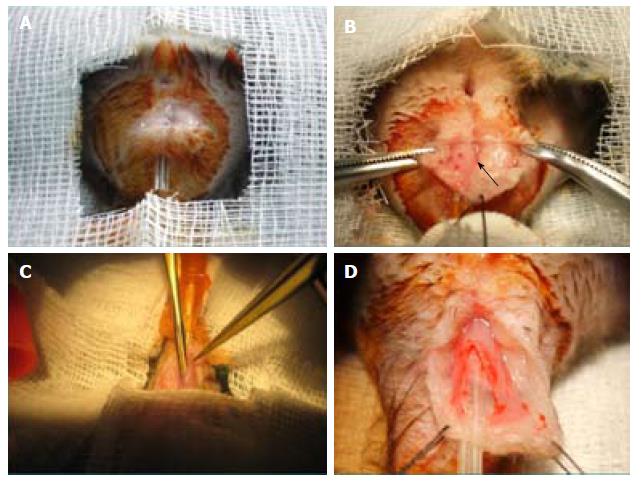
Figure 1 Sphincter anal injury model.
A: An arciform 10-mm to 15-mm anterior perianal incision (3 mm to 5 mm from the anal verge) was performed; B: Adipose tissue was dissected sideways to expose 20 mm of conduit until visualization of the thin, darker line of the external sphincter was achieved (arrow); C: An incision was made in the muscular layer until submucosa herniation, then submucosal dissection progressed in a longitudinal fashion, downward (until the anal verge or near the marking stitch) and upward (until 10 mm had been completed); D: Final aspect of sphincter section.
- Citation: Trébol J, Georgiev-Hristov T, Vega-Clemente L, García-Gómez I, Carabias-Orgaz A, García-Arranz M, García-Olmo D. Rat model of anal sphincter injury and two approaches for stem cell administration. World J Stem Cells 2018; 10(1): 1-14
- URL: https://www.wjgnet.com/1948-0210/full/v10/i1/1.htm
- DOI: https://dx.doi.org/10.4252/wjsc.v10.i1.1


