Copyright
©The Author(s) 2003.
World J Gastroenterol. Sep 15, 2003; 9(9): 2068-2072
Published online Sep 15, 2003. doi: 10.3748/wjg.v9.i9.2068
Published online Sep 15, 2003. doi: 10.3748/wjg.v9.i9.2068
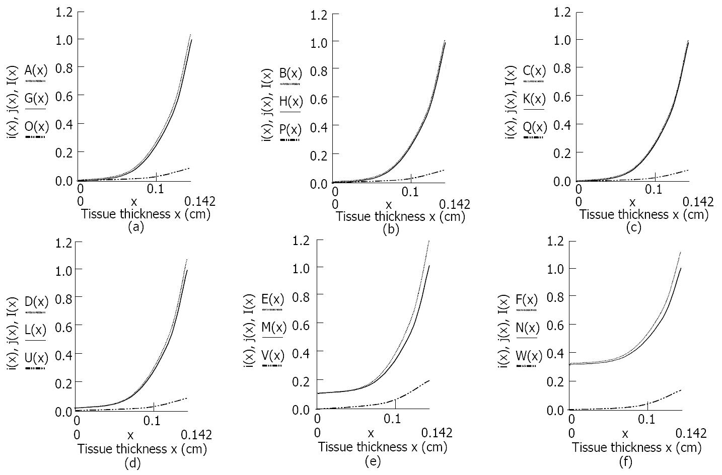
Figure 5 Light distribution of i (x), j (x), I (x) of human normal small intestine tissue in Kubelka-Munk two-flux model changed with tissue thickness at six different wavelengths of laser radiation.
(a) A(x), G(x) and O(x) respectively represented the forward and backward, and the total scattered photon fluxes of the tissue at 476.5 nm laser irradiation. (b) B(x), H(x) and P(x) respectively represented the forward, backward and the total scattered photon fluxes of the tissue at 488 nm laser irradiation. (c) C(x), K(x) and Q(x) respectively represented the forward, backward and the total scattered photon fluxes of the tissue at 496.5 nm laser irradiation. (d) D(x), L(x) and U(x) respectively represented the forward, backward and the total scattered photon fluxes of the tissue at 514.5 nm laser irradiation. (e) E(x), M(x) and V(x) respectively represented the forward, backward and the total scattered photon fluxes of the tissue at 532 nm laser irradiation. (f) F(x), N(x) and W(x) respectively represented the forward, backward and the total scattered photon fluxes of the tissue at 808 nm laser irradiation.
-
Citation: Wei HJ, Xing D, Wu GY, Jin Y, Gu HM. Optical properties of human normal small intestine tissue determined by Kubelka-Munk method
in vitro . World J Gastroenterol 2003; 9(9): 2068-2072 - URL: https://www.wjgnet.com/1007-9327/full/v9/i9/2068.htm
- DOI: https://dx.doi.org/10.3748/wjg.v9.i9.2068


