Copyright
©The Author(s) 2003.
World J Gastroenterol. Sep 15, 2003; 9(9): 2060-2064
Published online Sep 15, 2003. doi: 10.3748/wjg.v9.i9.2060
Published online Sep 15, 2003. doi: 10.3748/wjg.v9.i9.2060
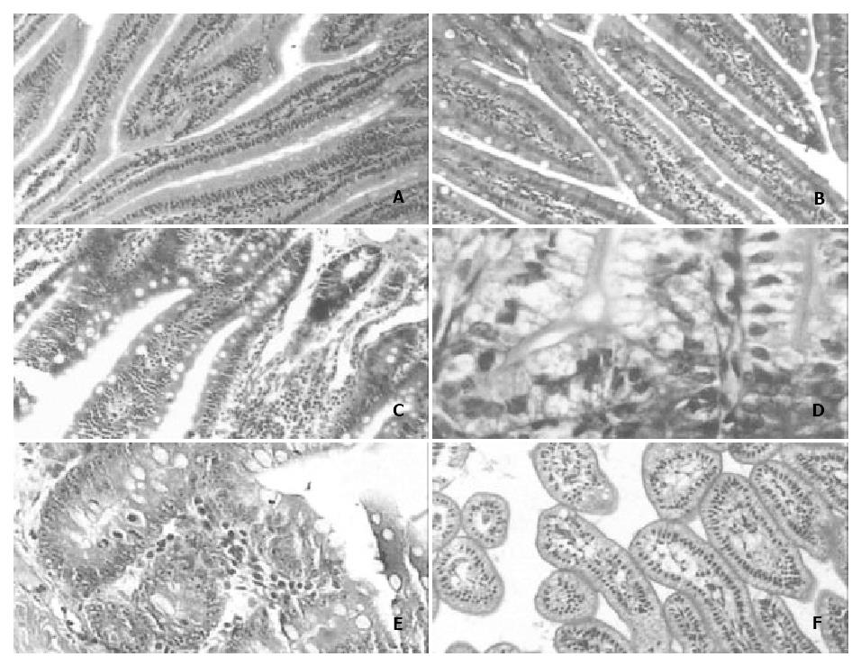
Figure 1 A: The number of goblet cells and the morphology of jejunal villi in the postnatal 3rd month × 100.
B: The number of goblet cells in rat jejunal villi in the postnatal 3rd month × 100. C: The number of goblet cells and the morphology of jejunal villi in the postnatal 12th month × 100. D: There were few or no goblet cells in the jejunal glands of newborn rats × 400; E: The number of goblet cells in rat jejunal gland was much higher in the postnatal 12th month than in any other month × 400. F: The jejunal villi of newborn rats displayed ateliosis and were very small × 200.
- Citation: Wang L, Li J, Li Q, Zhang J, Duan XL. Morphological changes of cell proliferation and apoptosis in rat jejunal mucosa at different ages. World J Gastroenterol 2003; 9(9): 2060-2064
- URL: https://www.wjgnet.com/1007-9327/full/v9/i9/2060.htm
- DOI: https://dx.doi.org/10.3748/wjg.v9.i9.2060


