Copyright
©The Author(s) 2003.
World J Gastroenterol. Sep 15, 2003; 9(9): 2036-2039
Published online Sep 15, 2003. doi: 10.3748/wjg.v9.i9.2036
Published online Sep 15, 2003. doi: 10.3748/wjg.v9.i9.2036
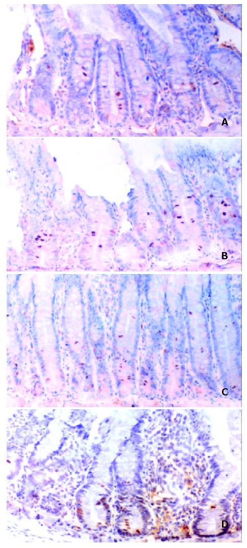
Figure 1 The expression of phosphorylated p38 MAPK in control group (A), ischemia group (B), ischemia-reperfusion 6 h (C) and 12 h (D) groups.
The positive expression of p38 MAPK signals was localized mainly in the lower half of the crypts and in the cytoplasm of the crypt cells. The positively stained cells increased remarkably after 6 h, and reached their peak at 12 h after reperfusion, which was about 35.6% of the total cells in crypts. At this stage, the positive staining was primarily localized in nucleus of crypt cells.
- Citation: Fu XB, Xing F, Yang YH, Sun TZ, Guo BC. Activation of phosphorylating-p38 mitogen-activated protein kinase and its relationship with localization of intestinal stem cells in rats after ischemia-reperfusion injury. World J Gastroenterol 2003; 9(9): 2036-2039
- URL: https://www.wjgnet.com/1007-9327/full/v9/i9/2036.htm
- DOI: https://dx.doi.org/10.3748/wjg.v9.i9.2036


