Copyright
©The Author(s) 2003.
World J Gastroenterol. Sep 15, 2003; 9(9): 1915-1919
Published online Sep 15, 2003. doi: 10.3748/wjg.v9.i9.1915
Published online Sep 15, 2003. doi: 10.3748/wjg.v9.i9.1915
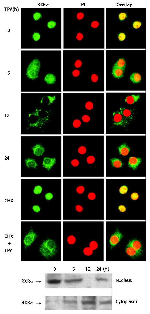
Figure 2 Translocation of RXRα from the nucleus to the cyto-plasm induced by TPA.
Cells were treated with TPA for differ-ent time periods or CHX (10 g/L) for 3 hrs as required. (A) Cells were immunostained with anti-RXRα antibody followed by corresponding FITC-conjugated anti-IgG secondary anti-body to show RXRα protein. Simultaneously, cells were stained with PI to display the nucleus. The fluorescent image was ob-served under laser-scanning confocal microscope. (B) Nuclear and cytoplasmic fractions were prepared as described in the Materials and Methods. RXRα was revealed by Western blot.
- Citation: Ye XF, Liu S, Wu Q, Lin XF, Zhang B, Wu JF, Zhang MQ, Su WJ. Degradation of retinoid X receptor α by TPA through proteasome pathway in gastric cancer cells. World J Gastroenterol 2003; 9(9): 1915-1919
- URL: https://www.wjgnet.com/1007-9327/full/v9/i9/1915.htm
- DOI: https://dx.doi.org/10.3748/wjg.v9.i9.1915


