Copyright
©The Author(s) 2003.
World J Gastroenterol. Aug 15, 2003; 9(8): 1799-1803
Published online Aug 15, 2003. doi: 10.3748/wjg.v9.i8.1799
Published online Aug 15, 2003. doi: 10.3748/wjg.v9.i8.1799
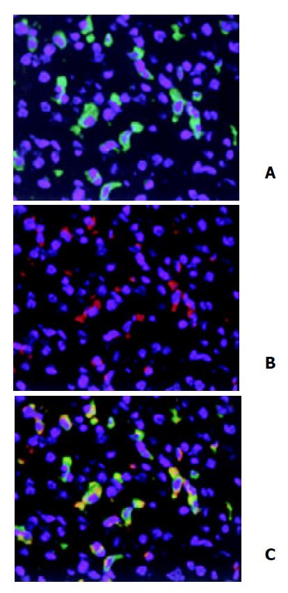
Figure 2 Double immunofluorescent staining of HO-1 local-ization in liver.
After three doses of LPS treatment, HO-1 and CD68 in liver tissue were detected by double immunofluores-cent staining with polyclonal rabbit antibody against HO-1 followed by FITC-conjugated anti-rabbit IgG (green) and mono-clonal rat antibody against mouse CD68 followed by Cy3-con-jugated anti-rat IgG (red). Cell nuclei were counterstained with bis-benzimide (blue). A: HO-1; B: Macrophages (Kupffer cells); C: HO-1 + Macrophages. magnification × 400.
- Citation: Song Y, Shi Y, Ao LH, Harken AH, Meng XZ. TLR4 mediates LPS-induced HO-1 expression in mouse liver: Role of TNF-α and IL-1β. World J Gastroenterol 2003; 9(8): 1799-1803
- URL: https://www.wjgnet.com/1007-9327/full/v9/i8/1799.htm
- DOI: https://dx.doi.org/10.3748/wjg.v9.i8.1799


