Copyright
©The Author(s) 2003.
World J Gastroenterol. Aug 15, 2003; 9(8): 1667-1674
Published online Aug 15, 2003. doi: 10.3748/wjg.v9.i8.1667
Published online Aug 15, 2003. doi: 10.3748/wjg.v9.i8.1667
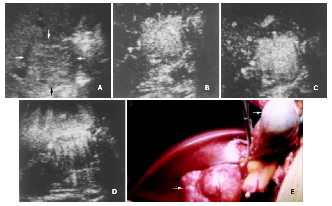
Figure 5 A 29-year-old man with focal nodular hyperplasia.
A. Intercostal section of precontrast conventional sonography exhibits an isoechoic lesion (arrows) in segment V of the liver with a diameter of 4.8 cm. B-D. Contrast-enhanced C-cube gray scale sonography at 49 s (B) and 60 s (C) after injection of Levovist shows that intranodular signals enhance earlier than the liver parenchyma, and enhancement sustains in the portal venous phase until the delayed phase (140 s, D), suggestive of slow wash-out of contrast in the lesion. E. Appearance of the lesion and its surrounding organs in laparotomy. The lesion (arrow) protrudes from the liver with a smooth capsule while the gallbladder (arrowhead) is lifted with hemostatic forceps. Histopathology of the tumor revealed focal nodular hyperplasia.
- Citation: Wang WP, Ding H, Qi Q, Mao F, Xu ZZ, Kudo M. Characterization of focal hepatic lesions with contrast-enhanced C-cube gray scale ultrasonography. World J Gastroenterol 2003; 9(8): 1667-1674
- URL: https://www.wjgnet.com/1007-9327/full/v9/i8/1667.htm
- DOI: https://dx.doi.org/10.3748/wjg.v9.i8.1667


